Category: Ataxia
Objective: This study aimed to investigate eye movements, specifically dysmetric saccades and cerebellar nystagmus, in Spinocerebellar Ataxia Type-12 (SCA12) using functional magnetic resonance imaging (fMRI) to assess oculomotor abnormalities and their association with cerebellar dysfunction.
Background: SCA12, caused by a CAG repeat expansion in the PPP2R2B gene on chromosome 5, is a rare autosomal dominant ataxia primarily found in the Indian subcontinent [1]. It is characterized by gait ataxia, dysarthria, tremor, and saccadic abnormalities [2]. However, limited research has explored saccadic velocity and dysmetria in SCA12.
Method: Forty genetically confirmed SCA12 patients (mean age: 52.67±8.94 years) and 25 healthy controls (mean age: 49.36±8.16 years) were recruited (Table 1). Participants underwent ICARS, MMSE, and cognitive tests before fMRI scanning on a 3T Philips scanner. A 3D T1-weighted Turbo Field Echo (TFE) sequence was used with the following parameters: TR = 8.1 ms, TE = 3.7 ms, FOV = 250 mm, 1 mm isovoxel, 350 slices (thickness = 0.5 mm), flip angle = 10°. Task-based functional MRI (fMRI) data included 180 dynamics across 35 contiguous axial slices with parameters TR = 2000 ms and TE = 40 ms. Simultaneous task-based fMRI paradigm and eye-tracking data acquisition were implemented using SuperLab software (v5.0, Cedrus Corporation, USA) and MR-compatible binocular LCD goggles (Nordic Neuro Lab, Norway) with an eye tracker (Arrington Research Inc., USA). Data analysis was conducted using SPM and CONN software. A saccadic paradigm (Figure 1), comprising rest and active blocks was presented to elicit horizontal, vertical, and circular saccades around a central fixation point.
Results: SCA12 patients exhibited slower saccades, reduced pupil reflex, and lower cognitive scores (Figure 2). Functional connectivity analysis revealed inhibitory connectivity from cerebellar regions to dorsal attention and sensorimotor networks during circular saccades. In horizontal tasks, posterior cerebellum-to-right intraparietal sulcus connectivity was suppressed, while facilitatory connectivity from the cerebellum to visual nodes was observed. Up-down saccades showed reduced cerebellum-to-frontoparietal connectivity (Figure 3 and 4).
Conclusion: SCA12 patients exhibit abnormal oculomotor patterns and altered cerebellar connectivity, suggesting compensatory mechanisms to counteract cerebellar dysfunction [3,4].
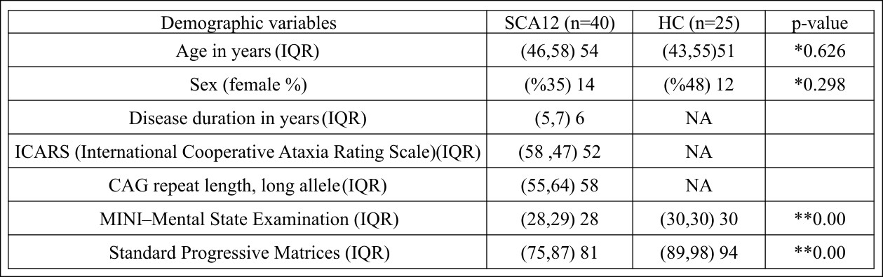
Demographic details in SCA12 and HC
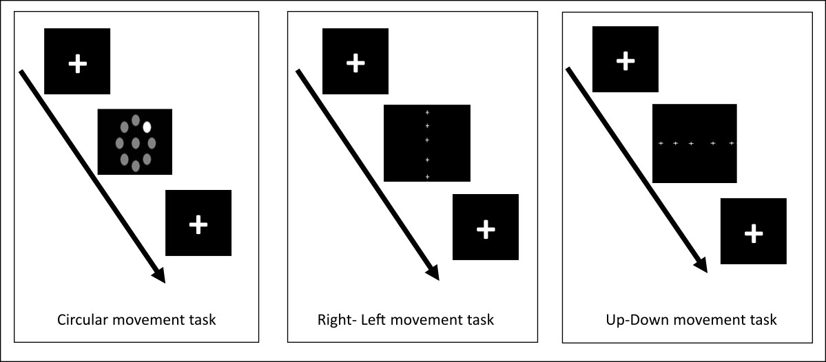
Schematic overview of the eye movement tasks
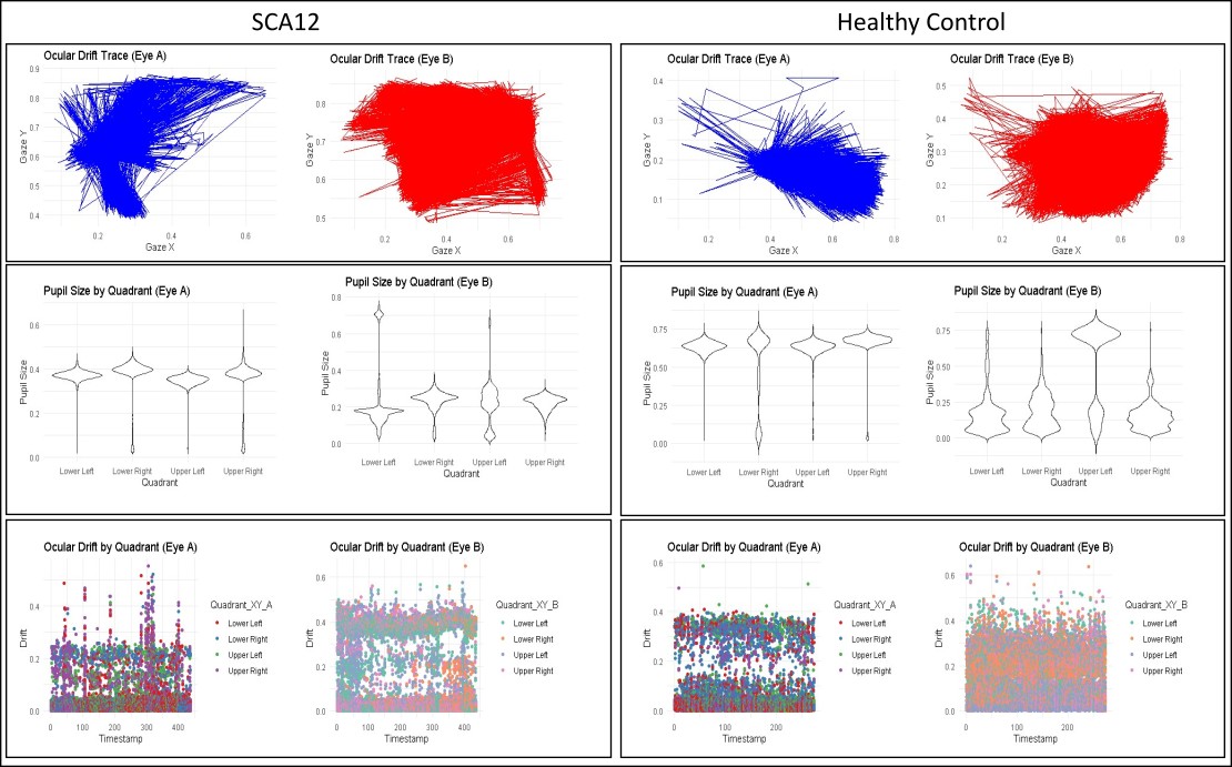
Eye-tracking data collected using binocular
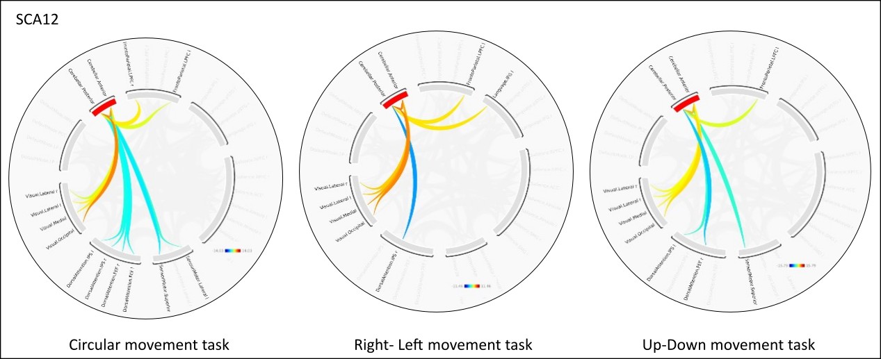
ROI-to-ROI functional connectivity in the SCA12
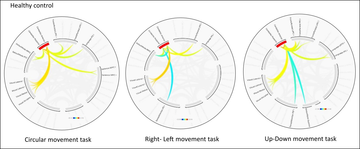
ROI-to-ROI functional connectivity in the healthy
References: 1. Holmes SE, Hearn EO, Ross CA, Margolis RL. SCA12: an unusual mutation leads to an unusual spinocerebellar ataxia. Brain Res Bull. 2001 Nov;56(3–4):397–403.
2. Rosini F, Pretegiani E, Battisti C, Dotti MT, Federico A, Rufa A. Eye movement changes in autosomal dominant spinocerebellar ataxias. Neurological Sciences. 2020 Jul 4;41(7):1719–34.
3. Stoodley CJ. The cerebellum and cognition: Evidence from functional imaging studies. In: Cerebellum. 2012. p. 352–65.
4. Yang Z, Zhong S, Carass A, Ying SH, Prince JL. Deep Learning for Cerebellar Ataxia Classification and Functional Score Regression. In 2014. p. 68–76.
To cite this abstract in AMA style:
P. Pankaj, A. Srivastava, S. Kumaran, A. Garg, R. Agarwal, M. Kumar, A. Sonakar, A. Nehra. Oculomotor Abnormalities in Spinocerebellar Ataxia Type-12: A Functional Neuroimaging Study [abstract]. Mov Disord. 2025; 40 (suppl 1). https://www.mdsabstracts.org/abstract/oculomotor-abnormalities-in-spinocerebellar-ataxia-type-12-a-functional-neuroimaging-study/. Accessed July 10, 2026.« Back to 2025 International Congress
MDS Abstracts - https://www.mdsabstracts.org/abstract/oculomotor-abnormalities-in-spinocerebellar-ataxia-type-12-a-functional-neuroimaging-study/