Category: Choreas (Non-Huntington's Disease)
Objective: To analyze the clinical profile of patients with acute chorea at a Brazilian tertiary hospital emergency room (ER).
Background: Chorea involves involuntary, rapid movements that flow randomly between body parts and can arise from different causes. (1) While chronic chorea is well understood, acute cases need urgent evaluation to find treatable causes. Recognizing clinical and imaging patterns aids in accurate diagnosis and management. (2,3)
Method: A prospective observational cohort study evaluated patients with acute chorea in the ER of Hospital Geral de Fortaleza, Brazil. Data on demographics, symptoms, comorbidities, medication history, neuroimaging findings, and diagnostic outcomes were collected from January 2024 to March 2025.
Results: We screened 3,077 patients during this period, including eleven cases with chorea (0.36%). Two were excluded due to chronic symptom onset, leaving nine patients (77.8% female) for analysis (Figure 1). Median age was 58 years (range, 9-89). Hemichorea was the predominant pattern in 77.8% of cases, while generalized chorea was present in 22.2%.
Common comorbidities included diabetes and hypertension (36.4% each), with one patient having HIV. Chorea etiologies were vascular (44.4%), metabolic (33.3%), autoimmune (9.1%), and infectious (9.1%) (Table 1). Brain MRI revealed basal ganglia abnormalities in 66.7% of cases. In three cases, basal nuclei were structurally normal. Among hemichorea patients (n=7), one had symptoms on the same side as lesion.
The main treatment was haloperidol with clonazepam. For resistant cases, clozapine and amantadine were added. Insulin was given for all cases of diabetic striatopathy. One patient received immunosuppressive therapy (MIPV plus cyclophosphamide) and another started combined antiretroviral therapy with antimicrobials for neurotoxoplasmosis. Most patients improved, but one needed extended management due to persistence movements.
Conclusion: Our findings indicate that acute chorea is a rare condition, accounting for less than 1% of cases seen in the emergency department. It is linked to various causes, with vascular and metabolic issues being the primary contributors, as shown in previous series (3). Stroke and diabetic striatopathy were the most common underlying diagnoses in our cohort. Recognizing the correlations between clinical presentations and imaging results can enhance early diagnosis and help guide appropriate treatment interventions.
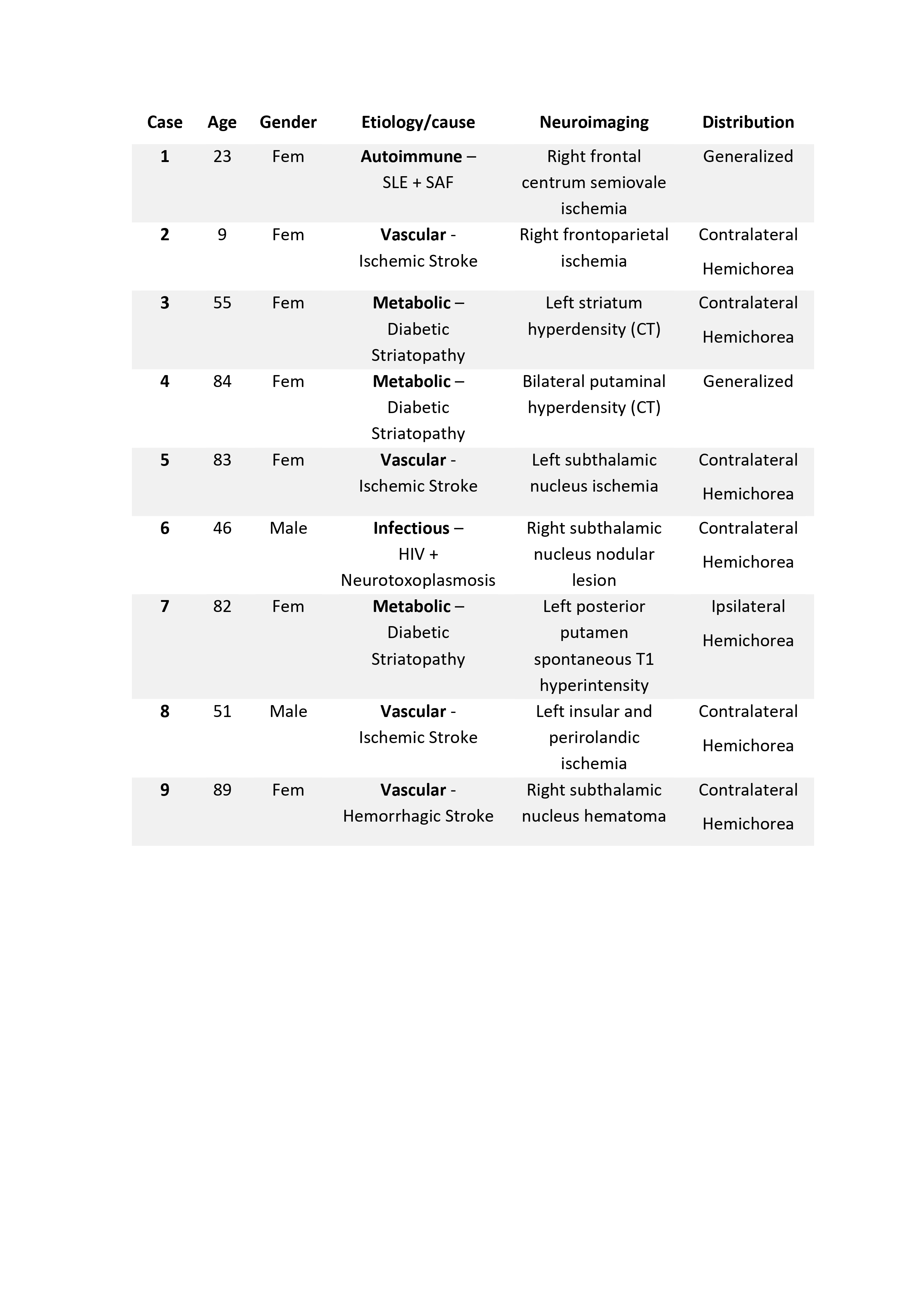
Table 1 – Acute chorea patients profile (N=9).
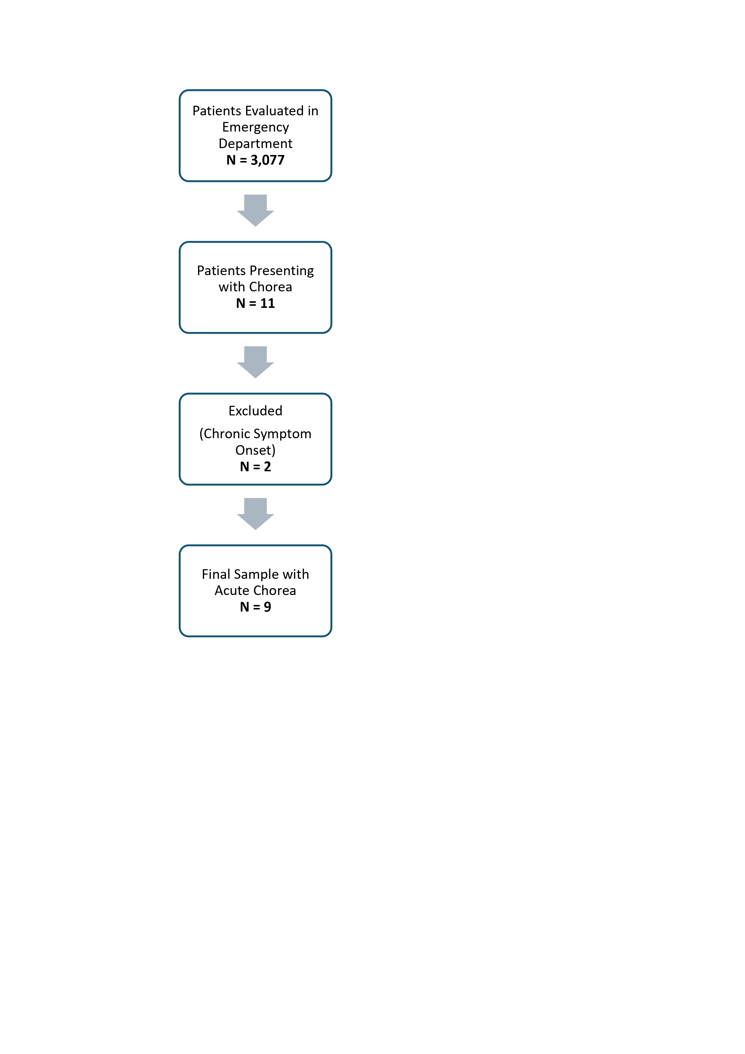
Figure 1: Patient inclusion flowchart.
References: 1.Jankovic,J.,Hallett,M.,Okun,M.S.,Comella,C.,Fahn,S.,&Goldman,J.(2021).Principles and Practice of Movement Disorders. Elsevier. https://doi.org/10.1016/B978-0-323-31071-0.01001-5
2. Cardoso F. (2021). Acute chorea in adults: old and new truths. Arquivos de neuro-psiquiatria, 79(3), 187–188. https://doi.org/10.1590/0004-282X-ANP-2021-E003
3.Silva, G. D., Parmera, J. B., & Haddad, M. S. (2021). Acute chorea: case series from the emergency room of a Brazilian tertiary-level center. Arquivos de neuro-psiquiatria, 79(3), 233–237. https://doi.org/10.1590/0004-282X-ANP-2020-0124
To cite this abstract in AMA style:
AS. Lima Verde, F. Rolim, A. Catunda, AR. Marinho, F. Carvalho. Acute Chorea in the Emergency Department: A Prospective Cohort Study [abstract]. Mov Disord. 2025; 40 (suppl 1). https://www.mdsabstracts.org/abstract/acute-chorea-in-the-emergency-department-a-prospective-cohort-study/. Accessed July 8, 2026.« Back to 2025 International Congress
MDS Abstracts - https://www.mdsabstracts.org/abstract/acute-chorea-in-the-emergency-department-a-prospective-cohort-study/