Objective: The purpose of this study was to explore shear wave elastography (SWE)’s ability to identify task-specific dystonic muscles by determining if muscle stiffness increased post exercise in suspected primary dystonic, but not in control muscles.
Background: Identifying dystonic muscles in lower-limb, task-specific dystonia (TSD) is crucial for optimizing therapeutic interventions. However, the two primary tools used for muscle identification have significant limitations. Computerized motion analysis is time-consuming, costly, and often unavailable. Bedside electromyography is invasive and less reliable as audible muscle overactivity abates when the provocative tasks cease. Ultrasonic SWE, a clinically available tool, has the potential to accurately identify dystonic muscle activity by providing non-invasive, real-time, quantifiable stiffness measures in anatomical structures. SWE is standard practice in identifying pathologies in oncology and hepatology; more recently, its utility in monitoring muscle stiffness in spasmodic torticollis and spasticity has shown promise.
Method: We enrolled 8 participants with adult-onset, TSD, involving lower limb muscles, who were either toxin-naïve or >12 weeks post-injection. Participants were evaluated by our team to select a primary dystonic muscle based on affected limb’s phenomenology. For comparison, a contralateral control muscle was selected. SWE was collected pre-exercise (after 15 minutes of rest) and immediately post-exercise (dystonic symptoms for three minutes). Mean stiffness, represented by shear wave velocity (m/s), was calculated from six SWE measures captured per muscle. A Student’s t-test was used for comparisons.
Results: Stiffness increased 36% pre-to-post-exercise in the primary dystonic muscles (paired t-test, p=0.0036) while there was no change in stiffness in the control muscles (paired t-test, p=0.9485). The dystonic muscle demonstrated 15400% greater change in stiffness pre-to-post exercise, relative to the control muscle (2-sample t-test p=0.0015).
Conclusion: This study provides novel evidence that SWE can distinguish dystonic and non-dystonic muscle by quantifying the increase (or lack thereof) in stiffness pre-to-post exercise. Thus, in conjunction with clinical evaluation, SWE has potential as a biomarker for dystonic muscle identification assisting in refining targeted therapy in complex gait disorders.
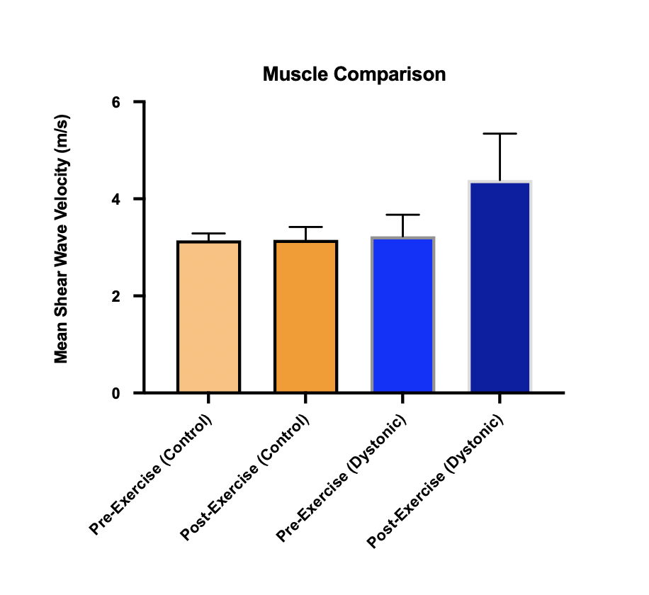
SWE Control vs Dystonic
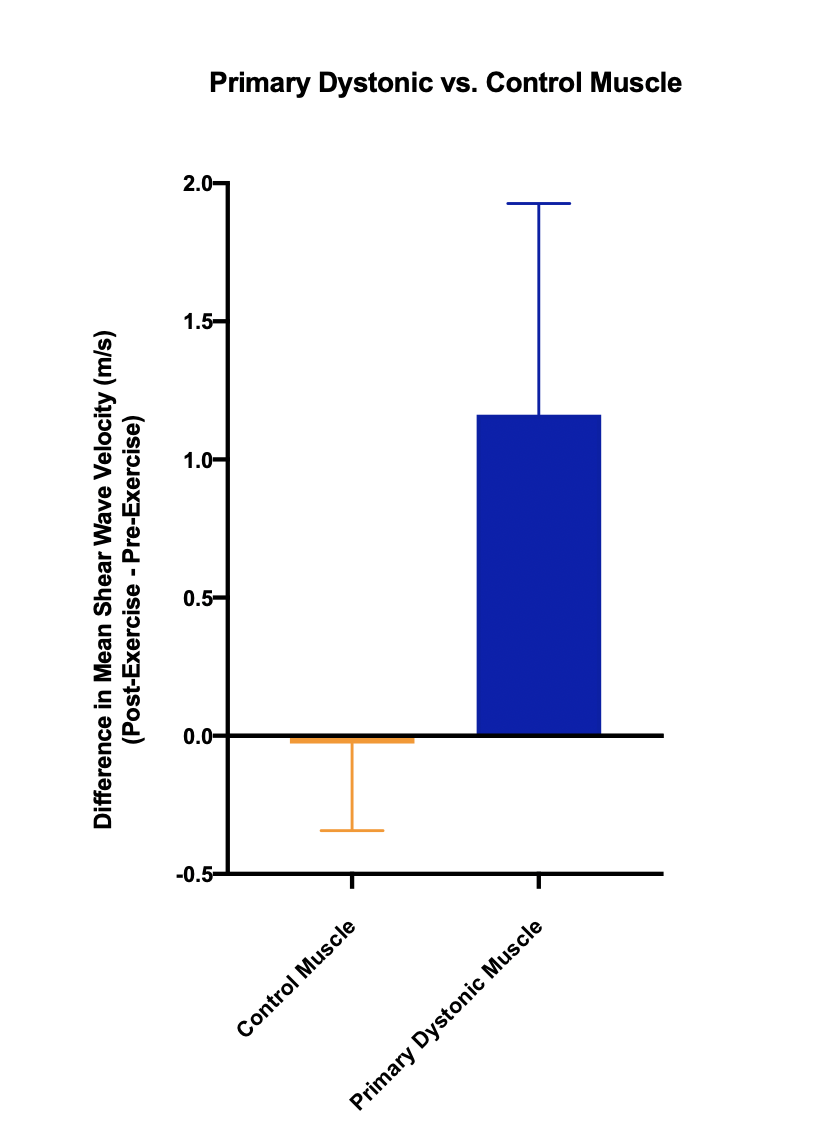
SWE Mean Difference Control vs Dystonic
References: Taljanovic MS, Gimber LH, Becker GW, et al. Shear-Wave Elastography: Basic Physics and Musculoskeletal Applications. Radiographics. 2017;37(3):855-870. doi:10.1148/rg.2017160116
Barr RG. Shear wave liver elastography. Abdom Radiol (NY). 2018;43(4):800-807. doi:10.1007/s00261-017-1375-1
Evans A, Whelehan P, Thomson K, et al. Quantitative shear wave ultrasound elastography: initial experience in solid breast masses. Breast Cancer Res. 2010;12(6):R104. doi:10.1186/bcr2787
Song Y, Zhang TJ, Li Y, Gao Y. Application of real-time shear wave elastography in the assessment of torsional cervical dystonia. Quant Imaging Med Surg. 2019;9(4):662-670. doi:10.21037/qims.2019.04.08
Hasegawa Y, Niimi M, Hara T, et al. Shear Wave Velocity to Evaluate the Effect of Botulinum Toxin on Post-Stroke Spasticity of the Lower Limb. Toxins (Basel). 2022;15(1):14. Published 2022 Dec 26. doi:10.3390/toxins15010014
To cite this abstract in AMA style:
A. Ly, A. Mushtaheed, A. Roberts, M. Druckenbrod, J. Stowers, M. Soliman, P. Lind, Y. Assefa, C. Stanley, A. Gravunder, J. Matsubara, C. Zampieri-Gallagher, D. Ehrlich, B. Karp, F. Gavelli, K. Alter. Pilot Study: Shear Wave Elastography as a Potential Biomarker in Task-Specific Dystonia [abstract]. Mov Disord. 2025; 40 (suppl 1). https://www.mdsabstracts.org/abstract/pilot-study-shear-wave-elastography-as-a-potential-biomarker-in-task-specific-dystonia/. Accessed July 10, 2026.« Back to 2025 International Congress
MDS Abstracts - https://www.mdsabstracts.org/abstract/pilot-study-shear-wave-elastography-as-a-potential-biomarker-in-task-specific-dystonia/