Category: Parkinson's Disease (Other)
Objective: To compared the effects of theta burst transcranial ultrasound stimulation (tbTUS) applied to M1, the GPi, and simultaneous M1 and GPi stimulation to investigate their respective therapeutic potentials
Background: Parkinson’s disease (PD) involves disruptions in the basal ganglia-thalamocortical circuitry, leading to a variety of motor dysfunctions. Low-intensity transcranial ultrasound stimulation (TUS) is a safe and non-invasive technique for stimulating the cortical and subcortical brain nuclei with high spatial precision.
Method: We employed a 2-channel transducer (500 kHz) for primary motor cortex (M1) stimulation and a 4-channel transducer (500 kHz) for internal globus pallidus (GPi) stimulation. Targeting of M1 and GPi were based on the motor hot spots and individual MRI, respectively. Brainsight neuronavigation system and BabelBrain were used for modeling and thermal simulation. We positioned both transducers over the corresponding M1 and GPi locations, simultaneously. Theta burst transcranial ultrasound stimulation (tbTUS) (duty cycle of 10%, pulse repetition frequency of 5 Hz) was then delivered for 120 seconds in three separate sessions under different conditions: 1) real stimulation of M1 with sham GPi stimulation, 2) real stimulation of GPi with sham M1 stimulation, and 3) simultaneous dual-site sonication. The left and right hemispheres were targeted sequentially. Movement Disorders Society Unified Parkinson Disease Rating Scale (MDS-UPDRS) part III scores and transcranial magnetic stimulation (TMS) measurements were recorded before and after tbTUS.
Results: We recruited 15 participants. Two participants dropped out of the study. Thirteen participants (mean age 62.6 ± 9.3 years and disease duration 7.5 ± 3.6 years) completed the study. We found that tbTUS of M1 significantly increased MEP ratio to baseline at T10 compared to tbTUS of GPi. Additionally, the mean stimulation intensity needed to elicit 1mV MEP increased after tbTUS of GPi at T10. Notably, MDS-UPDRS part III scores and bradykinesia subscores reduced after tbTUS of GPi at T30.
Conclusion: In this study, we demonstrated that tbTUS of M1 enhanced cortical excitability, while tbTUS of GPi tended to decreased cortical excitability in PD patients. Furthermore, tbTUS of GPi led to improvements in PD motor symptoms, particularly bradykinesia.
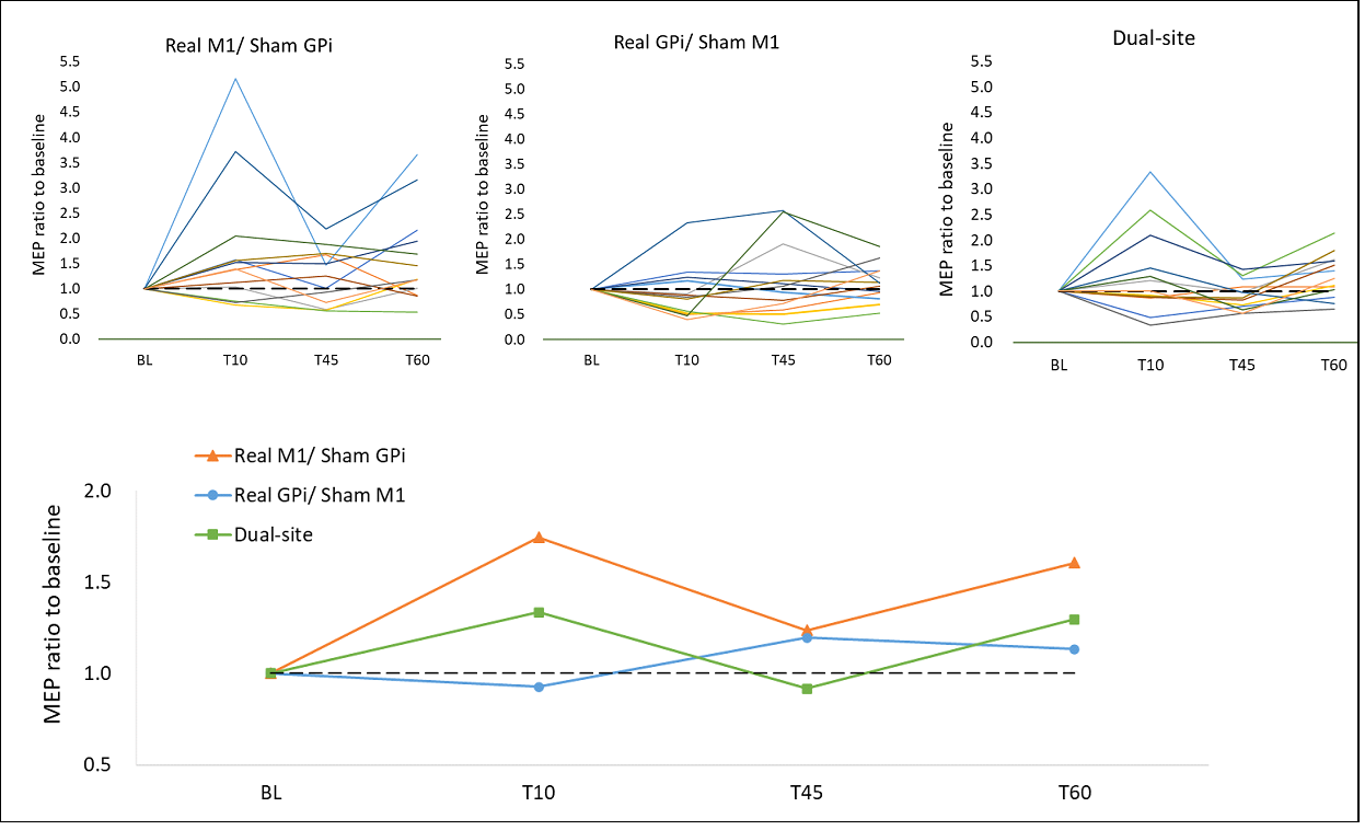
Changes of MEPs amplitude
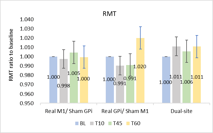
Change of resting motor threshold (RMT)
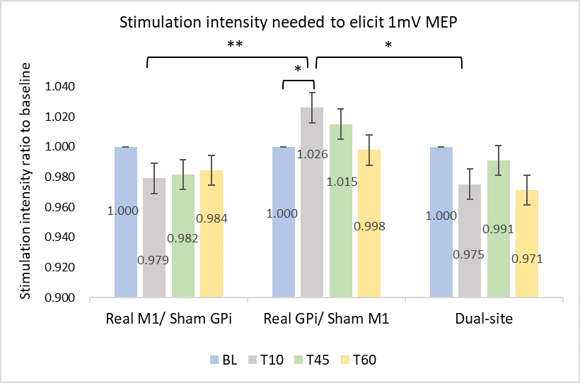
Changes of stimulation intensity
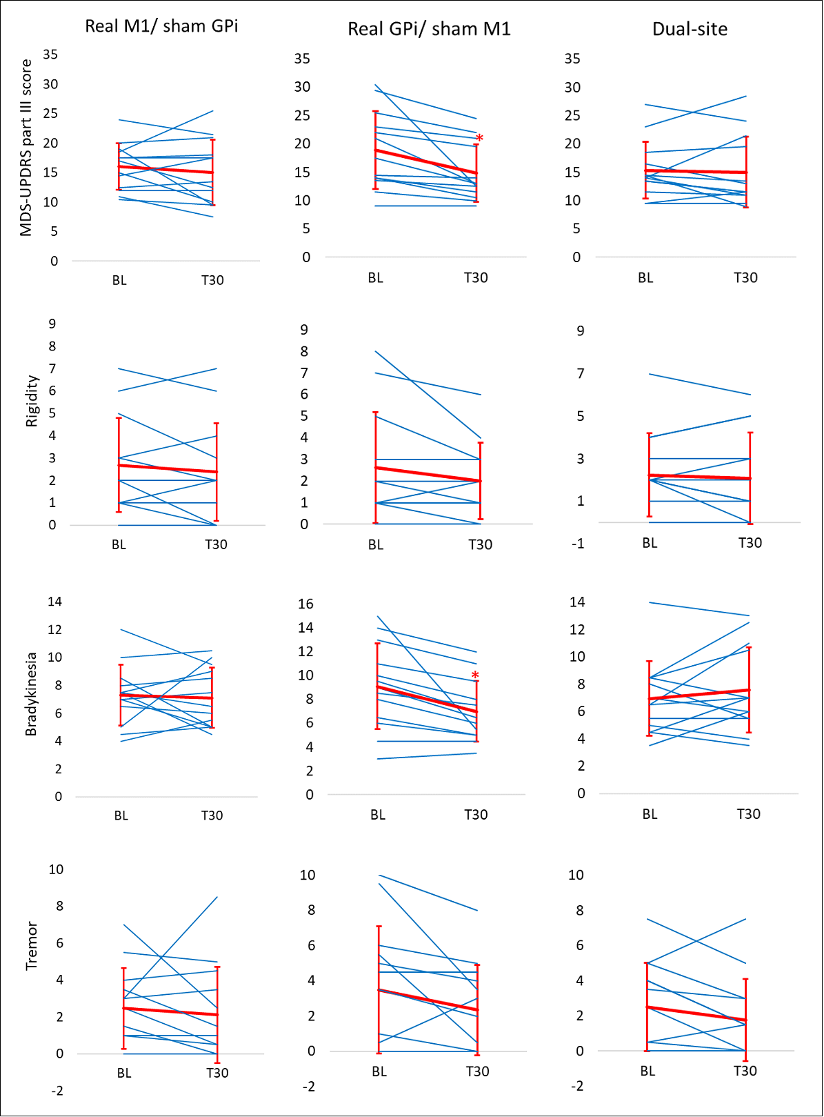
Changes of MDS-UPDRS part III scores and subscores
To cite this abstract in AMA style:
YY. Lin, N. Raies, T. Grippe, A. Bhattacharya, C. Sarica, G. Darmani, R. Chen. Theta burst transcranial ultrasound stimulation of internal globus pallidus and motor cortex in Parkinson’s disease [abstract]. Mov Disord. 2025; 40 (suppl 1). https://www.mdsabstracts.org/abstract/theta-burst-transcranial-ultrasound-stimulation-of-internal-globus-pallidus-and-motor-cortex-in-parkinsons-disease/. Accessed July 10, 2026.« Back to 2025 International Congress
MDS Abstracts - https://www.mdsabstracts.org/abstract/theta-burst-transcranial-ultrasound-stimulation-of-internal-globus-pallidus-and-motor-cortex-in-parkinsons-disease/