Category: Parkinson's disease: Neuroimaging
Objective: Our study examined brain network changes during the transition to Freezing of Gait (FOG) versus voluntary stopping. Identifying specific neural alterations may help establish reliable neurophysiological markers for predicting FOG, with potential therapeutic applications.
Background: Most Parkinson’s disease (PD) patients will experience FOG where there is an involuntary cessation of gait that often leads to falls. The changes in local brain network dynamics in real-time during the transition to FOG remain unexplored and are necessary to inform closed-loop therapeutic interventions. This study investigates the neural oscillation changes within local networks during transitions to FOG compared to voluntary stopping (VS) events using ambulatory EEG.
Method: Eighteen PD patients (69.6 ± 11.5 years) with varying FOG severity were assessed in their “Off” medication state. Motor measures (MDS-UPDRS III, Hoehn & Yahr) were recorded, and ambulatory EEG was acquired using a 32-channel BioSemi Active-Two system. Participants completed a Timed Up and Go (TUG) task and a voluntary stopping (VS) TUG task, halting upon a verbal “stop” command (Cao et al., 2021; Zampieri et al., 2010). Two trial types (FOG and VS) were analyzed, each comprising three 2-second epochs: Normal Walking (pre-transition), Transition (trFOG: before freezing; trVS: before voluntary stop), and FOG or VS. A total of 277 FOG and 51 VS trials were included (Fig 1A). Functional brain networks were estimated at the source level across four frequency bands. Graph theory-based modularity metrics were applied to compare network properties during transition states (trFOG/trVS) preceding FOG and VS events (Fig1B).
Results: The results revealed that networks during the trFOG exhibited increased local information processing (modularity) compared to the trVS periods. This was observed in the right frontoparietal (FPR) (p = 0.009, FDR), right visual (VISR) (p = 0.005, FDR) and insula (INS) (p = 0.007, FDR) networks within the beta frequency band (Fig 1C, 1D). Additionally, increased beta segregation was observed in VS compared to trVS within the FPR network (p = 0.016, FDR).
Conclusion: Our findings suggest that beta-driven local network reorganization may serve as an early marker for predicting FOG episodes. Real-time EEG dynamics could be leveraged for integration into sensing devices to guide future interventions.
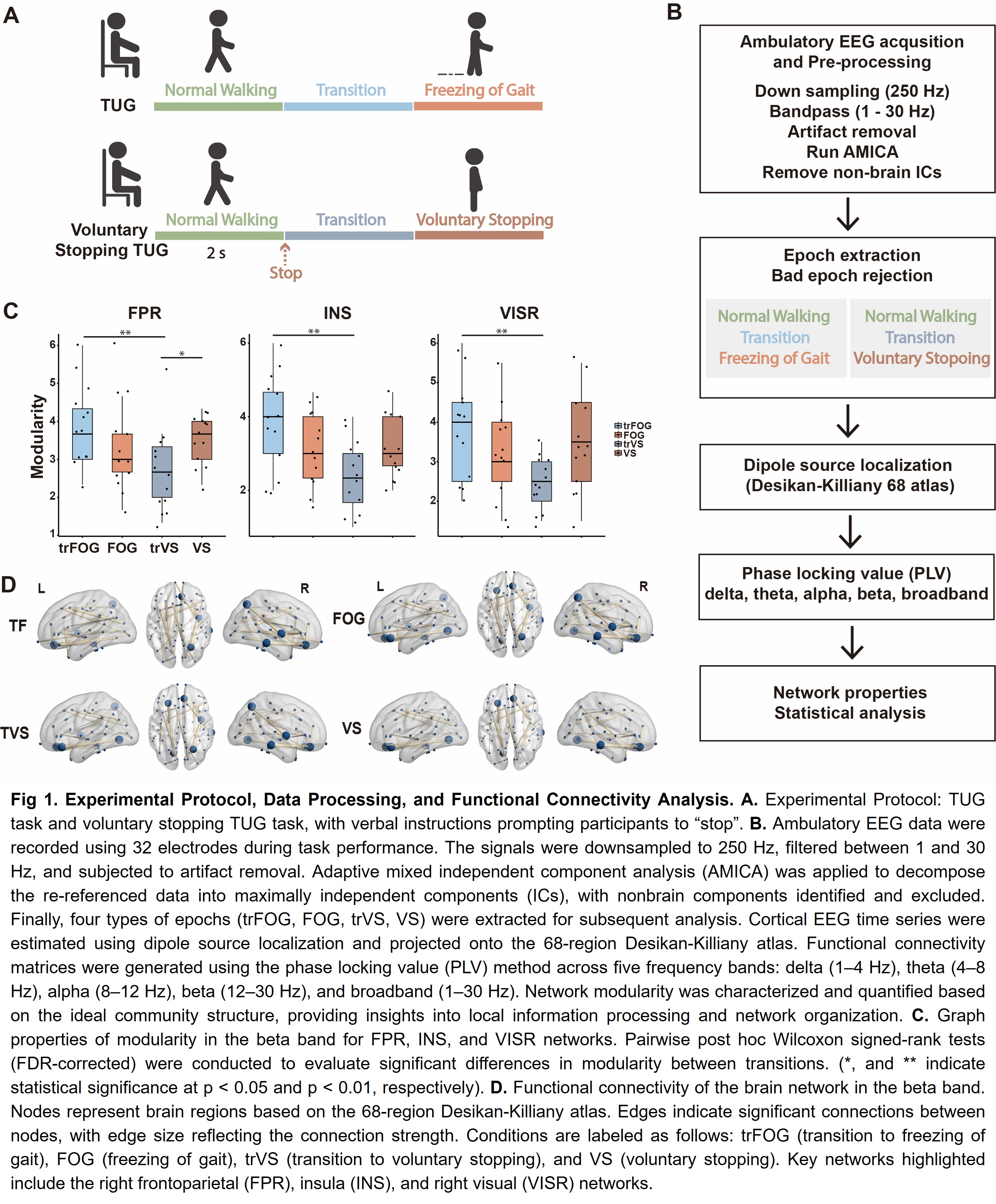
Fig 1. Experimental Protocol and Data Processing.
References: Cao, Z., John, A. R., Chen, H.-T., Martens, K. E., Georgiades, M., Gilat, M., . . . Engineering, R. (2021). Identification of EEG dynamics during freezing of gait and voluntary stopping in patients with Parkinson’s disease. 29, 1774-1783.
Zampieri, C., Salarian, A., Carlson-Kuhta, P., Aminian, K., Nutt, J. G., Horak, F. B. J. J. o. N., Neurosurgery, & Psychiatry. (2010). The instrumented timed up and go test: potential outcome measure for disease modifying therapies in Parkinson’s disease. 81(2), 171-176.
To cite this abstract in AMA style:
Y. Tian, E. Matar, S. Lewis. Utilising Beta Power in Local Networks to Predict Freezing of Gait Events in Parkinson’s Disease [abstract]. Mov Disord. 2025; 40 (suppl 1). https://www.mdsabstracts.org/abstract/utilising-beta-power-in-local-networks-to-predict-freezing-of-gait-events-in-parkinsons-disease/. Accessed July 10, 2026.« Back to 2025 International Congress
MDS Abstracts - https://www.mdsabstracts.org/abstract/utilising-beta-power-in-local-networks-to-predict-freezing-of-gait-events-in-parkinsons-disease/