Objective: The purpose of our study is to explore the differences in ReHo patterns under acute and chronic pain conditions in Parkinson’s disease (PD) patients, aiming to provide basis for the study of pain mechanism and the search of pain markers in PD.
Background: The impact of pain on the quality of life in PD is serious. The results of functional magnetic resonance imaging (fMRI) on patients with PD related pain are controversial, besides, whether the brain activity stimulated by acute pain is consistent with that by chronic pain in PD still needs further investigation.
Method: According to the inclusion and exclusion criteria, 75 PD patients and 30 healthy controls (HC) were included in experiment, and the patients were divided into two groups according to the pain score. Basic data of subjects were collected, and evaluation of motor function, disease stage, cognition, emotion, and other symptoms were performed. Three groups of subjects underwent fMRI scanning and then data were statistically analyzed.
Patients with chronic pain or without pain underwent a second scan with a cold pressor test after the first scan, and then data were statistically analyzed.
Results: There was no statistical difference in basic data and disease characteristics among three groups (P>0.05). Analysis showed there were significant differences in middle frontal gyrus (MFG), superior frontal gyrus (SFG), postcentral gyrus, precentral gyrus, and superior parietal gyrus (GRF correction, voxel P<0.001, cluster P<0.05)[figure1]. In patients with chronic pain, Reho in above regions were significantly higher (P<0.05 or P<0.001) than those with no pain. In MFG/SFG (P<0.01), postcentral/precentral gyrus (P<0.001), Reho in patients with chronic pain were significantly higher than HC, but in superior parietal gyrus were lower (P<0.05) than HC. In patients with no pain, they had decreased Reho in superior parietal gyrus (P<0.001) compared with HC.
The altered brain regions in patients with chronic pain before and after stimulation included postcentral and precentral gyrus (P<0.001)[figure2], while in patients with no pain only included the middle temporal gyrus (P<0.001)[figure3].
Conclusion: Chronic and acute pain in PD have different but overlapping brain activation patterns, our results provide a basis for the study of the pathogenesis and imaging markers of acute and chronic pain in PD.
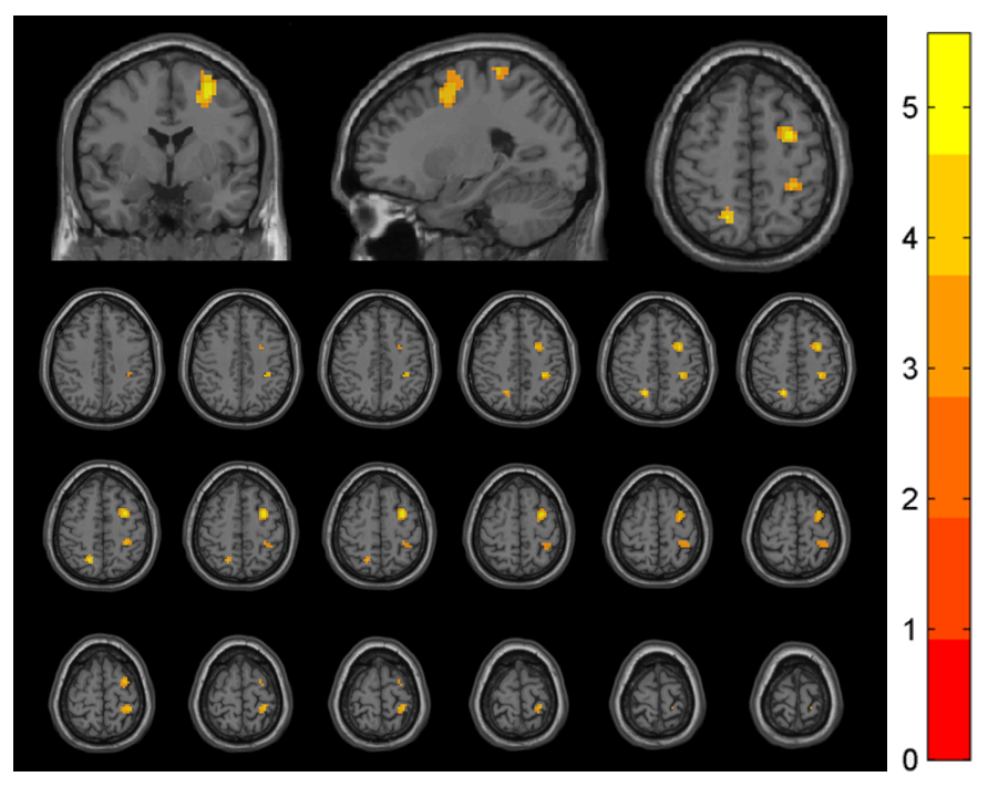
ReHo Differences in Brain Regions Across Groups
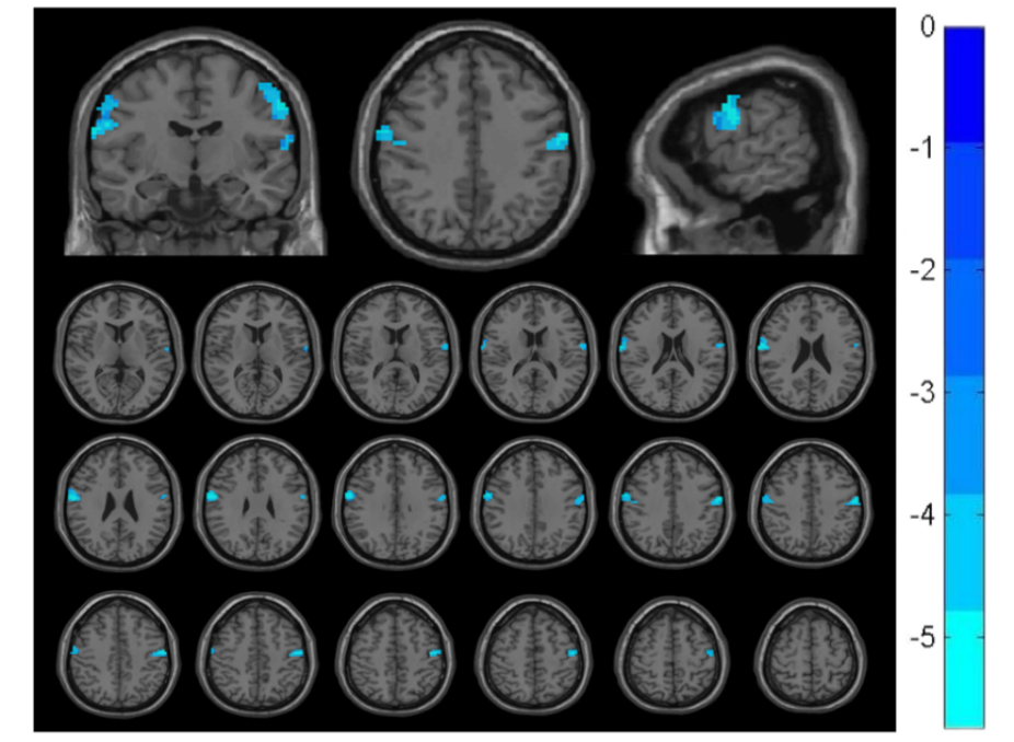
ReHo Changes in Pain Group Pre/Post-Stimulation
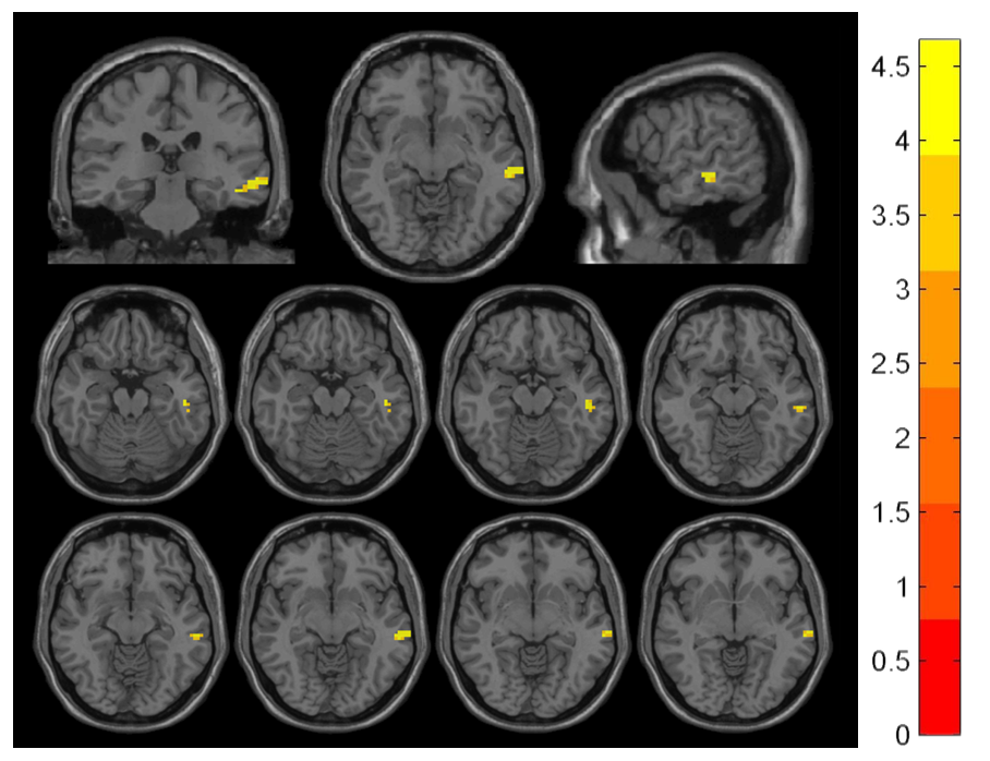
ReHo Changes in No Pain Group Pre/Post-Stimulation
To cite this abstract in AMA style:
CJ. Mao, JY. Wu, J. Cheng, YJ. Luo, QM. Jiang. A Resting-State fMRI Study of Regional Homogeneity about Pain in Parkinson’s Disease [abstract]. Mov Disord. 2025; 40 (suppl 1). https://www.mdsabstracts.org/abstract/a-resting-state-fmri-study-of-regional-homogeneity-about-pain-in-parkinsons-disease/. Accessed July 10, 2026.« Back to 2025 International Congress
MDS Abstracts - https://www.mdsabstracts.org/abstract/a-resting-state-fmri-study-of-regional-homogeneity-about-pain-in-parkinsons-disease/