Session Information
Date: Thursday, June 8, 2017
Session Title: Parkinson's Disease: Neuroimaging And Neurophysiology
Session Time: 1:15pm-2:45pm
Location: Exhibit Hall C
Objective: To explore the time varying brain connectivity networks in Parkinson’s disease (PD) during resting-state fMRI and to investigate the correlate between connectivity dynamics and performance on the clinical features.
Background: Most brain connectivity studies in PD are based on the stationary assumption where the brain connectivity networks are assumed to be time invariant. However, the dynamics of brain connectivity have been increasingly recognized as being important.
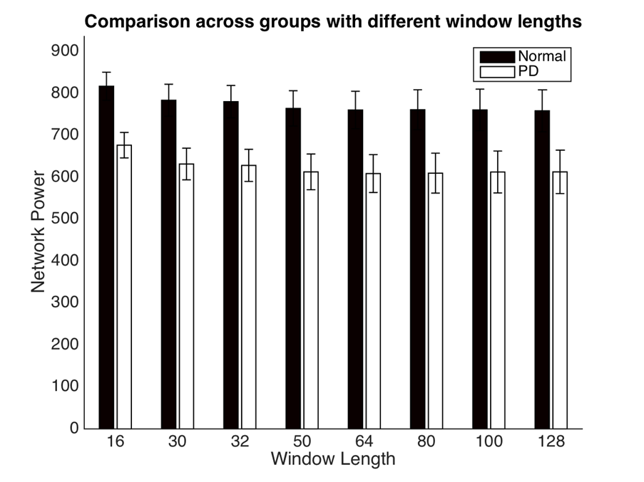
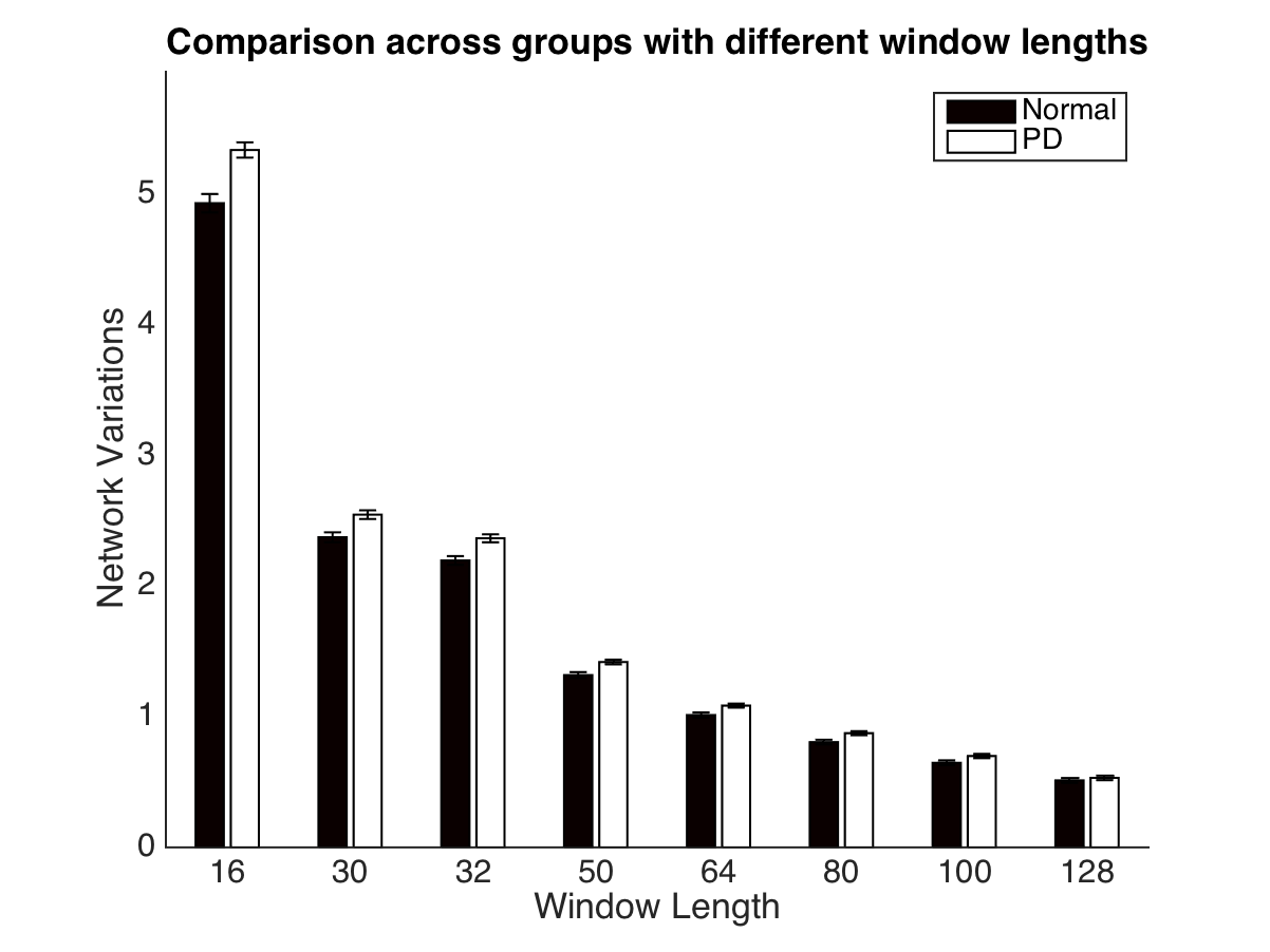
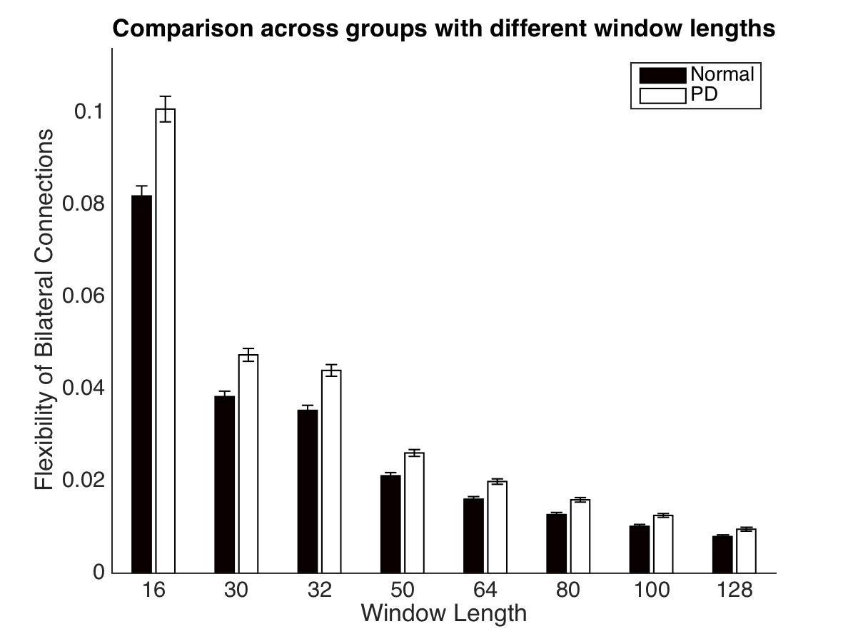
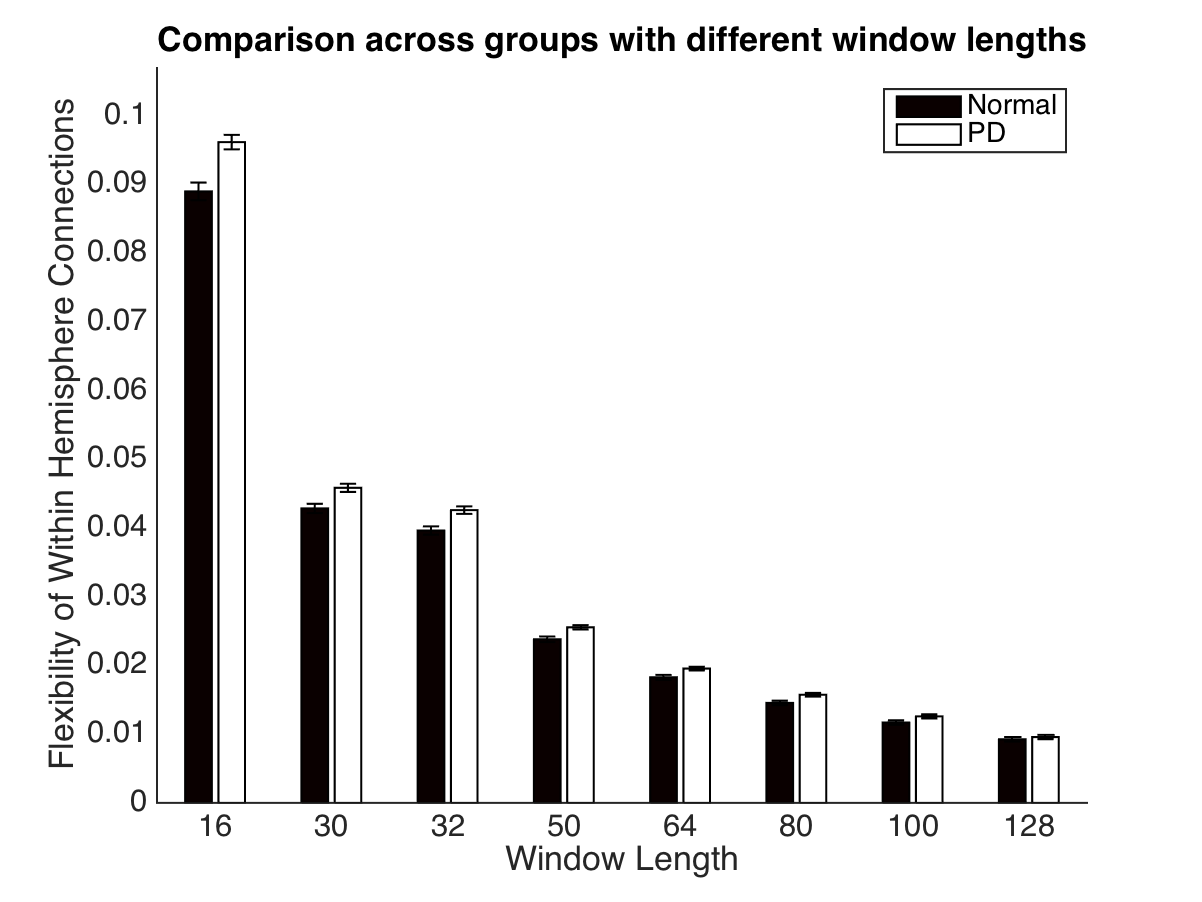
Methods: We compared the connectivity dynamics between PD groups (N=26) and normal control (N=24) with selected window length (16, 30, 32, 50, 64, 80, 100, 128 respectively) using sliding window strategy. The network variation and connectivity power were compared.
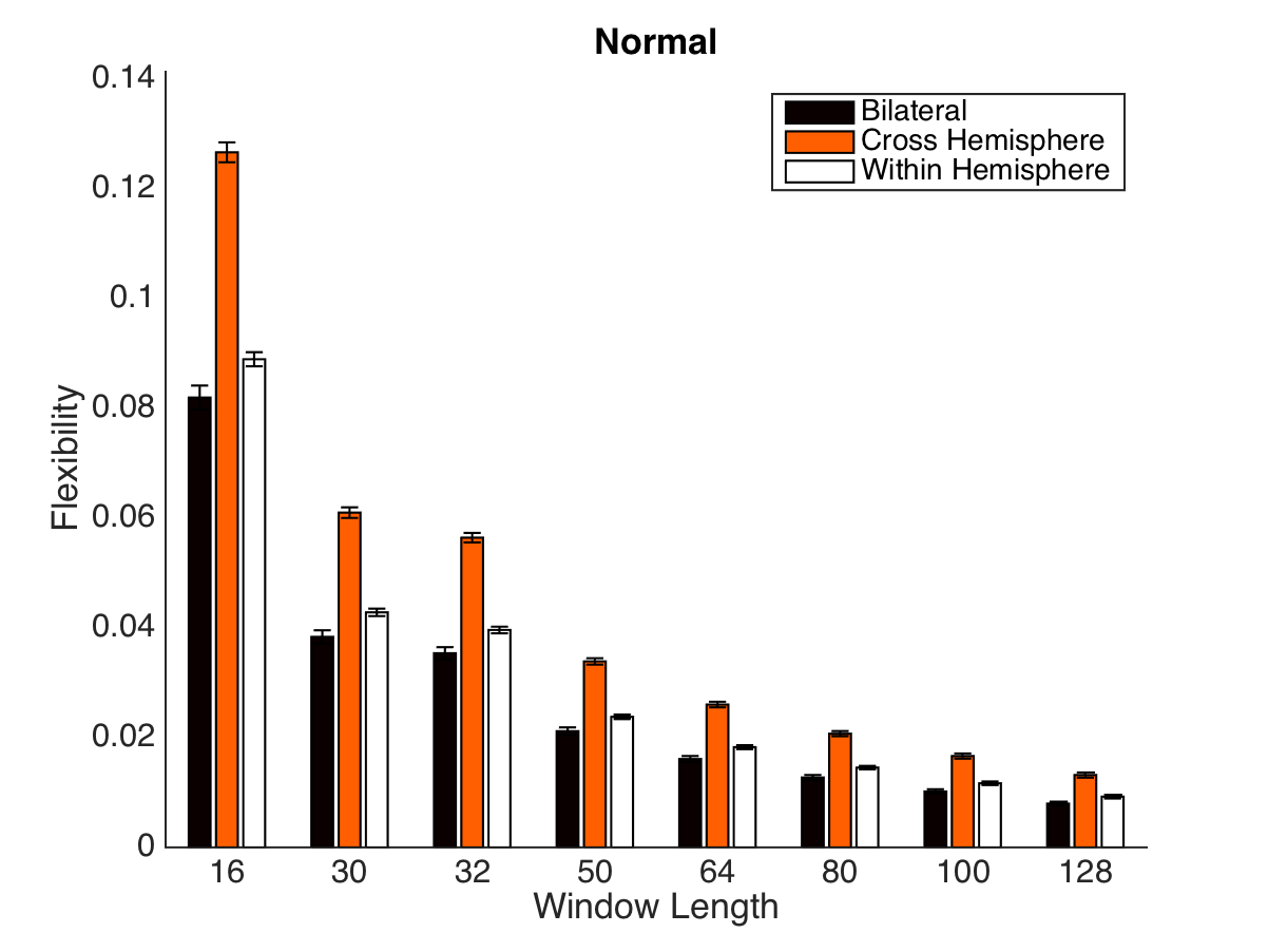
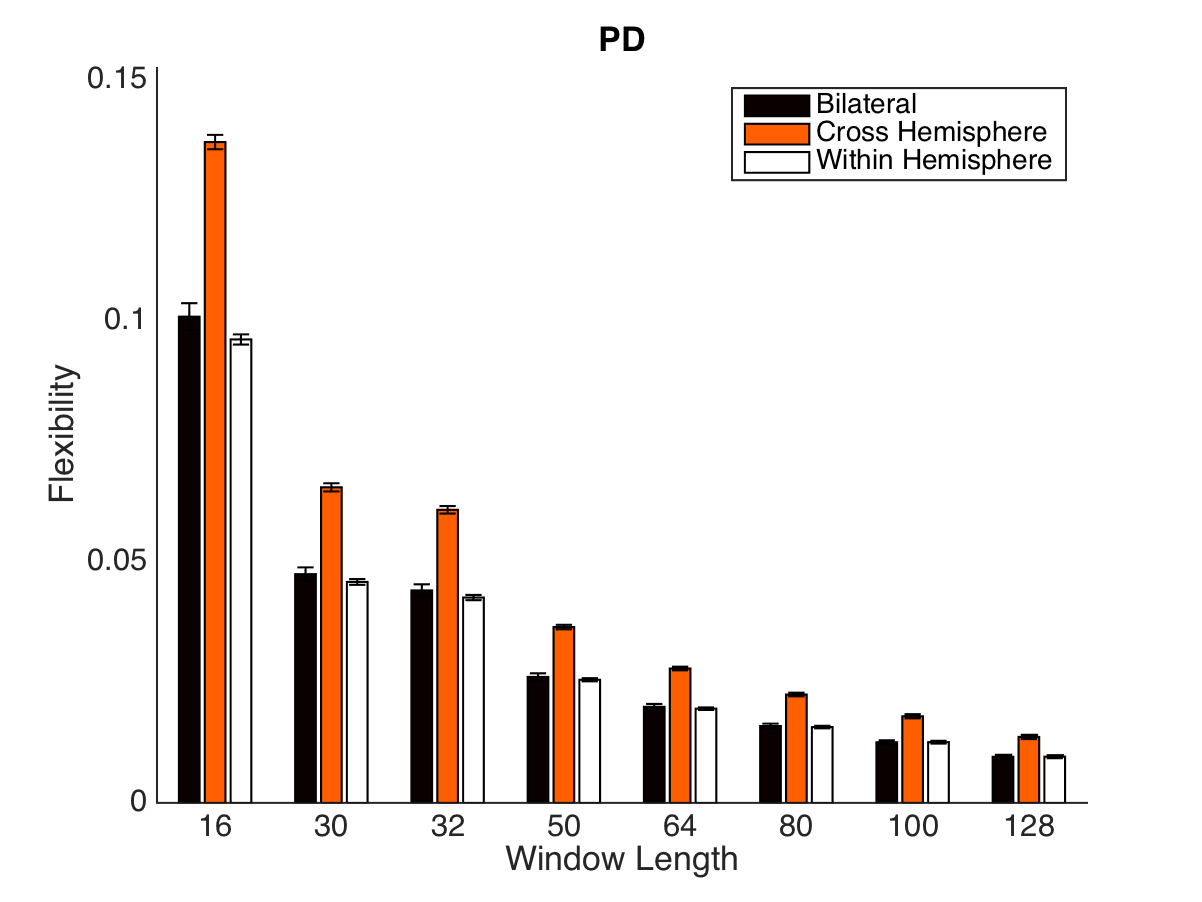
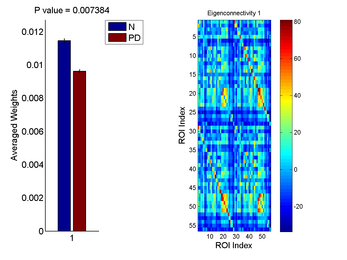
Results: The PD group demonstrated the lower connectivity power compared with that of normal group at all the window length.(Fig.1) The variations of brain connectivity was decreasing with the increasing of the window length (Fig.2). The bilateral connections have less variability while the cross hemisphere maintain higher variations(Fig.3,4). The differences between normal and PD groups has become less obvious with the increasing of the window length(Fig.5,6,7). At all the window length, the loading weights of first eigenconnectivity were correlated with the network dynamics(Fig.8). The age, gender and smoking were found to be correlated with the network power at all the window length.

Conclusions: The differences in flexibility revealed by brain time varying networks may further provide us the insights into brain function altered in PD.
To cite this abstract in AMA style:
X. Dan, A. Liu, J. Wang, M. McKeown, T. Wu, P. Chan. Alterations in Dynamic Brain Connectivity Patterns in Parkinson’s Disease [abstract]. Mov Disord. 2017; 32 (suppl 2). https://www.mdsabstracts.org/abstract/alterations-in-dynamic-brain-connectivity-patterns-in-parkinsons-disease/. Accessed July 5, 2026.« Back to 2017 International Congress
MDS Abstracts - https://www.mdsabstracts.org/abstract/alterations-in-dynamic-brain-connectivity-patterns-in-parkinsons-disease/