
Objective: To characterize the dopaminergic lesion pattern in behavioral variant frontotemporal dementia with parkinsonism (bvFTD-P) using dopamine transporter (DAT) positron emission tomography (PET), and to evaluate its potential value in differentiating bvFTD-P from other neurodegenerative parkinsonian syndromes.
Background: Behavioral variant of frontotemporal dementia (bvFTD) often presents with parkinsonism. Our study aimed to characterize the dopaminergic lesion pattern in bvFTD-parkinsonism (bvFTD-P) using dopamine transporter (DAT) positron emission tomography (PET) and to assess its potential value in differentiating bvFTD-P from progressive supranuclear palsy (PSP), multiple system atrophy–parkinsonism (MSA-P), and Parkinson’s disease (PD).
Method: Thirty bvFTD patients with parkinsonism were enrolled and underwent DAT PET imaging with either ¹¹C-CFT or 18F-FP-CIT. For comparison, 71 patients with PSP, 41 with MSA-P, 61 with late-onset PD (LOPD), and 43 healthy controls (HC) were included [figure 1]. DAT binding values in the caudate, anterior putamen, and posterior putamen were calculated, along with caudate/anterior putamen (C/AP) and caudate/posterior putamen (C/PP) ratios. Group differences were analyzed using generalized linear models. Receiver operating characteristic (ROC) analysis was performed to evaluate the diagnostic performance of DAT binding values and ratios.
Results: Compared with HC, patients with bvFTD-P showed significantly reduced DAT binding in the caudate, anterior putamen, and posterior putamen (all p < 0.01) [figure 2]. However, the pattern of impairment was caudate-predominant, reflected by lower C/AP and C/PP ratios [figure 3]. In contrast, PSP, MSA-P, and LOPD demonstrated a putamen-predominant DAT loss pattern. ROC analysis showed that DAT binding ratios, particularly the C/PP ratio, provided better discrimination between bvFTD-P and other groups than absolute DAT binding values. Visual assessment indicated that approximately two-thirds of bvFTD-P patients had markedly reduced DAT binding, while about one-third showed normal DAT scans.
Conclusion: A subset of bvFTD-P patients shows a caudate-predominant dopaminergic deficit pattern that differs from the putamen-dominant loss typically observed in PSP, MSA-P, and PD. DAT imaging, particularly striatal binding ratios such as C/PP, may serve as a useful imaging biomarker for differentiating bvFTD-P from other parkinsonian syndromes.
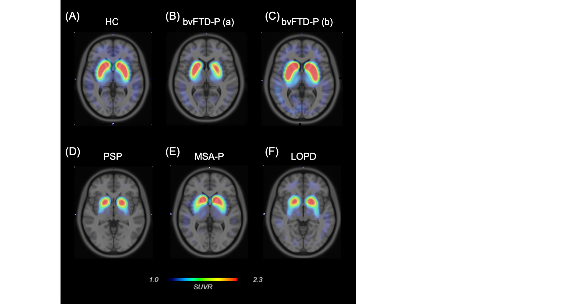
figure2
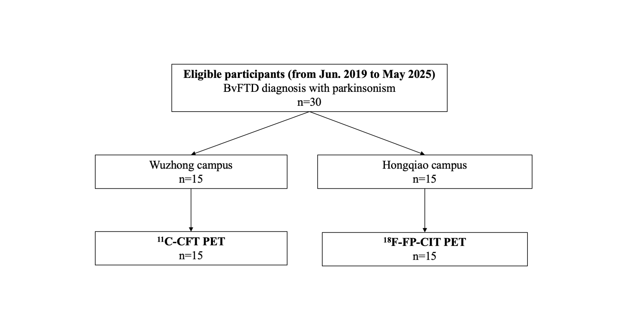
figure1
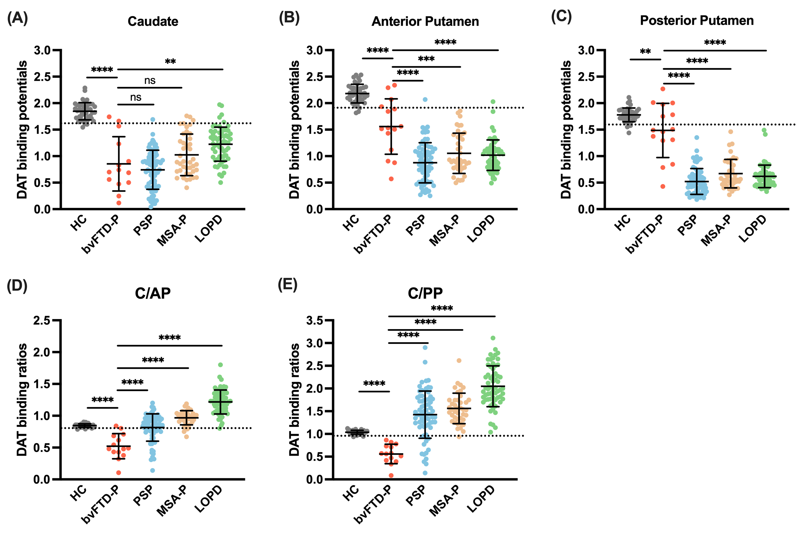
figure3
To cite this abstract in AMA style:
L. Feng, W. Liu, J. Wang, J. Wang, F. Liu. Caudate-Predominant Striatal Dopaminergic Deficits in bvFTD with Parkinsonism [abstract]. Mov Disord. 2026; 41 (suppl 1). https://www.mdsabstracts.org/abstract/caudate-predominant-striatal-dopaminergic-deficits-in-bvftd-with-parkinsonism/. Accessed October 1, 2026.« Back to 2026 International Congress
MDS Abstracts - https://www.mdsabstracts.org/abstract/caudate-predominant-striatal-dopaminergic-deficits-in-bvftd-with-parkinsonism/