Category: Parkinson's disease: Neuroimaging
Objective: Investigate the effects of exercise on resting-state (rs) fMRI functional connectivity (FC) in Parkinson’s Disease (PD).
Background: Recent studies have shown strong clinical evidence that exercise can attenuate disease progression in PD. However, the underlying cerebral mechanisms responsible remain unclear [1]. Understanding how exercise modulates FC in PD can provide valuable insights into the physiological underpinnings of this effect.
Method: 22 PD subjects and 21 age-matched healthy controls (HC) were enrolled in the study. A PD sub-group (N=7) was enrolled in a six-month exercise program: thrice weekly, supervised, 60-minute stationary cycling. An additional PD non-exercise control sub-group (N=9) was also followed over a six-month period. rs-fMRI data were collected for all subjects at baseline and after six-months.
Processing of the MRI data was done using FreeSurfer, and CONN. First level analysis, computed using subject specific ROI to ROI connectivity, determined the connection strengths between subcortical and motor regions which were hypothesized to be altered by both PD and exercise (left and right: anterior/middle/posterior putamen, caudate, precentral, postcentral, paracentral, superior-frontal, and cerebellum).
Second level analysis was performed separately to investigate the effects of the intervention (post vs. pre) and disease (PD vs. HC). To visualize exercise effects on disease altered connections, the changes in connection strengths over the six-month period (averaged across each sub-group) were regressed as a function of the differences in connection strengths due to disease (averaged across the PD and HC groups). To avoid the regression toward the mean effect, only those PD subject data not included in each correlation’s post vs. pre comparison were included in the corresponding PD vs. HC comparisons under the assumption of being representative of a wider sample.
Results: Changes in connectivity strength following the six-month intervention program were found to correlate with differences in connectivity strength due to PD in the exercise group only; with superior-frontal and cerebellum connections highlighted as important regions for this effect [figure1].
Conclusion: These data provide strong supporting evidence that exercise positively affects connectivity specifically impaired in PD and highlights regions most related to this effect.
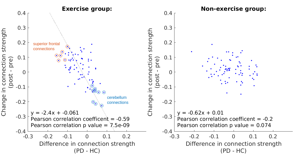
Effect of exercise program on PD rs-fMRI FC.
References: [1] M. E. Johansson et al., “Aerobic exercise alters brain function and structure in Parkinson’s disease: a randomized controlled trial,” Annals of neurology, 2022.
To cite this abstract in AMA style:
E. Reimers, C. Bevington, J. Mckenzie, S. Dhaliwal, J. Stoessl, V. Sossi. Effect of Exercise Intervention on Parkinson’s Disease Resting-State fMRI Functional Connectivity [abstract]. Mov Disord. 2025; 40 (suppl 1). https://www.mdsabstracts.org/abstract/effect-of-exercise-intervention-on-parkinsons-disease-resting-state-fmri-functional-connectivity/. Accessed April 7, 2026.« Back to 2025 International Congress
MDS Abstracts - https://www.mdsabstracts.org/abstract/effect-of-exercise-intervention-on-parkinsons-disease-resting-state-fmri-functional-connectivity/