Category: Parkinson's disease: Neuroimaging
Objective: To investigate correlations between local gyrification index (LGI) and neuropsychological variables in participants with Parkinson’s disease (PD) experiencing mild cognitive impairment (PD-MCI).
Background: PD is characterized by motor symptoms, but cognitive decline leading to PD-MCI affects approximately 50% of participants [1, 2]. Previous studies of PD-MCI have relied on conventional cortical measures (volume, area, thickness, curvature), which, while revealing cortical thinning, may not capture three-dimensional folding patterns. We hypothesized that LGI would identify distinct patterns that correlate with cognitive decline in PD-MCI [3, 4].
Method: Thirty participants were included: PD-MCI (n=18) and PD with normal cognition (PD-NC) (n=12). All participants’ data were collected using T1-weighted (T1w) sequences on a 3T Siemens Skyra scanner, along with neuropsychological (NP) assessments evaluating attention, working memory, language, memory, and visuospatial domains, as well as clinical measures. We employed FreeSurfer-7 to extract LGI of regions of interest using T1w MRI. Statistical analysis between LGI and NP variables was conducted using PALM in FSL and was considered significant at FDR pcorr<0.05.
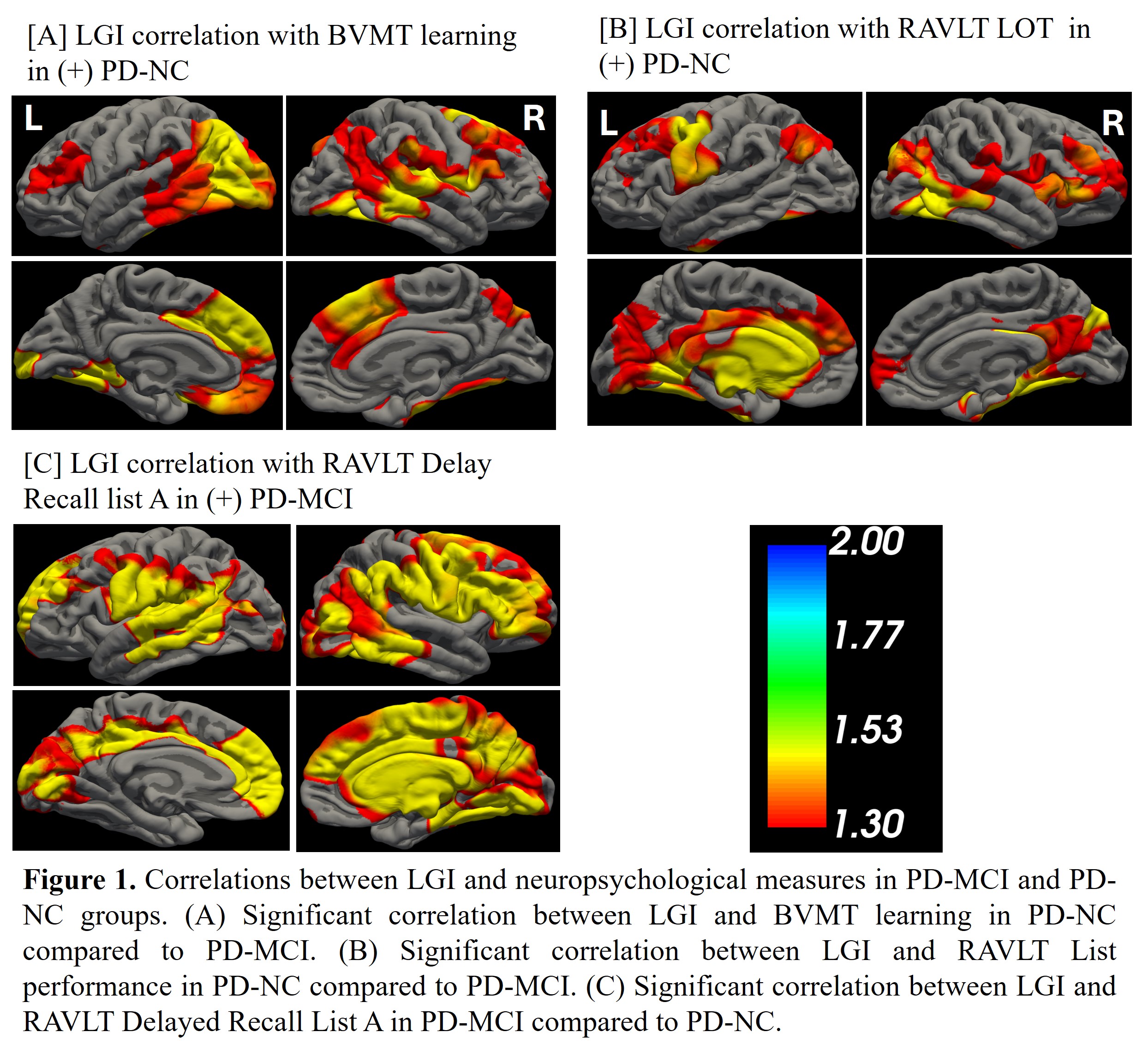
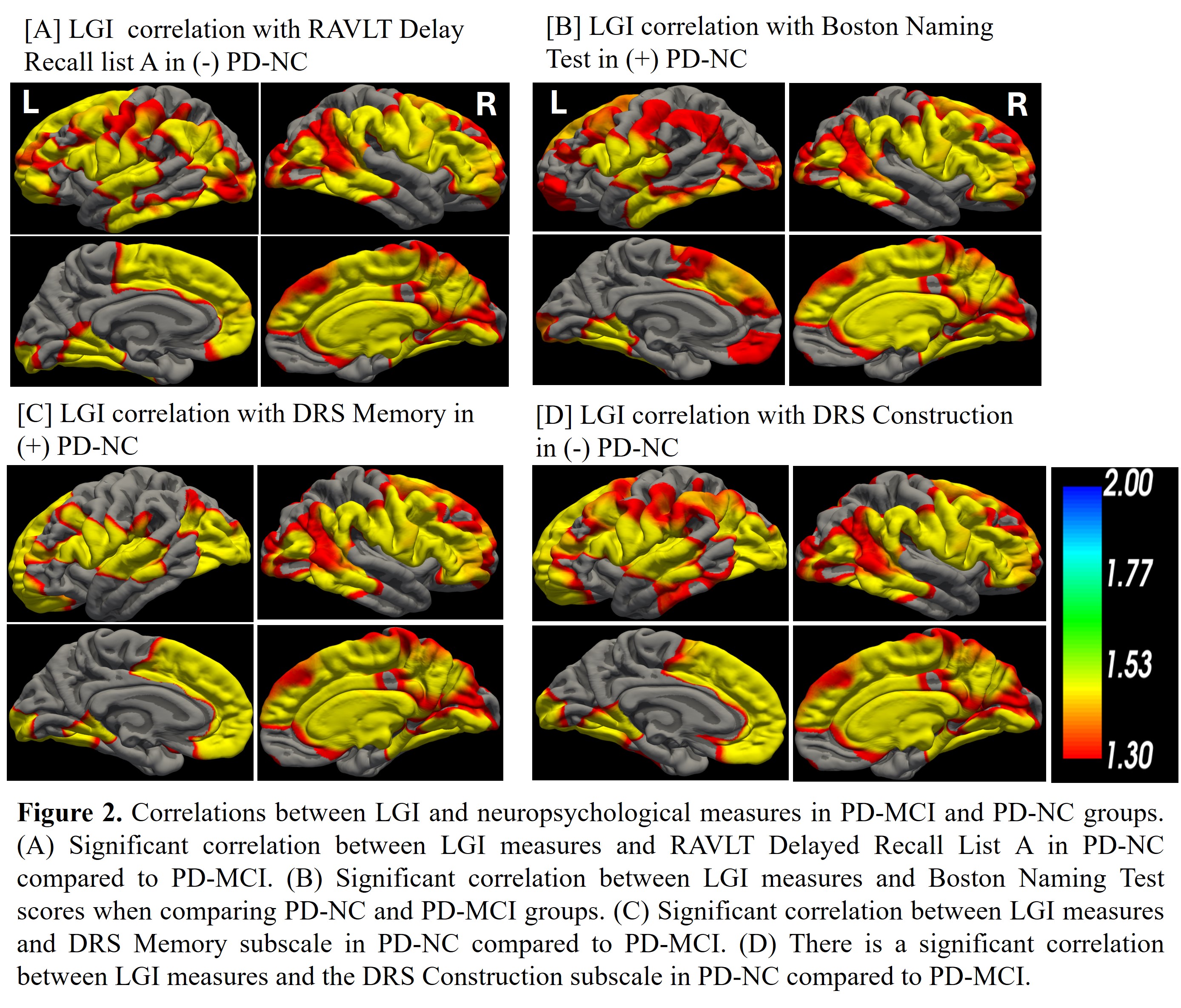
Results: Significant correlations between LGI measures and neuropsychological variables were observed in PD-MCI and PD-NC groups (precentral, postcentral, lingual, insula, temporal pole, medial-orbitofrontal, superior frontal, middle temporal, lateral occipital, etc.). PD-NC showed positive correlations between LGI and Brief Visuospatial Memory Test (BVMT) learning, Rey Auditory Verbal Learning Test (RAVLT) learning [Fig 1A-C], Dementia Rating Scale (DRS) memory, and Boston Naming Test. PD-MCI demonstrated a positive correlation between LGI and RAVLT delayed recall. Negative correlations were found in PD-NC between LGI and RAVLT delayed recall and DRS construction [Fig 2A-D].
Conclusion: Significant correlation patterns between LGI and cognitive performance were observed in both groups. Our study suggests that alterations in cortical gyrification may reflect distinct neuroanatomical substrates of cognitive impairment in PD. LGI analysis provides matching information to traditional cortical measures and may serve as a potential structural predictive marker for a better understanding of cognitive decline in PD.
Correlations: LGI and neuropsychological measures
Correlations: LGI and neuropsychological measures
References: [1] M. J. Armstrong and M. S. Okun, “Diagnosis and Treatment of Parkinson Disease: A Review,” JAMA, vol. 323, no. 6, pp. 548-560, Feb 11 2020, doi: 10.1001/jama.2019.22360.
[2] D. M. Cammisuli, S. M. Cammisuli, J. Fusi, F. Franzoni, and C. Pruneti, “Parkinson’s Disease-Mild Cognitive Impairment (PD-MCI): A Useful Summary of Update Knowledge,” Front Aging Neurosci, vol. 11, p. 303, 2019, doi: 10.3389/fnagi.2019.00303.
[3] Q. Shen et al., “Cortical gyrification pattern of depression in Parkinson’s disease: a neuroimaging marker for disease severity?,” Front Aging Neurosci, vol. 15, p. 1241516, 2023, doi: 10.3389/fnagi.2023.1241516.
[4] N. W. Sterling et al., “Stage-dependent loss of cortical gyrification as Parkinson disease “unfolds”,” Neurology, vol. 86, no. 12, pp. 1143-51, Mar 22 2016, doi: 10.1212/WNL.0000000000002492.
To cite this abstract in AMA style:
N. Chaurasiya, A. Katragadda, G. Rathi, J. Caldwell, Z. Mari, V. Mishra. Exploring local gyrification index to understand Parkinson’s disease with mild cognitive impairment [abstract]. Mov Disord. 2025; 40 (suppl 1). https://www.mdsabstracts.org/abstract/exploring-local-gyrification-index-to-understand-parkinsons-disease-with-mild-cognitive-impairment/. Accessed April 10, 2026.« Back to 2025 International Congress
MDS Abstracts - https://www.mdsabstracts.org/abstract/exploring-local-gyrification-index-to-understand-parkinsons-disease-with-mild-cognitive-impairment/