Category: Dystonia: Pathophysiology, Imaging
Objective: To report the case of a patient diagnosed with a solitary fibrous tumor located on the foramen magnum that presented with hand dystonia
Background: The foramen magnum solitary fibrous tumor (SFT) is one of the most rare and challenging tumors among all the SFT, the indolent development at the craniocervical junction makes clinical diagnosis complex and often leads to a large dimension tumor at diagnosis.
Method: Case report accompanied by video and neuroimaging.
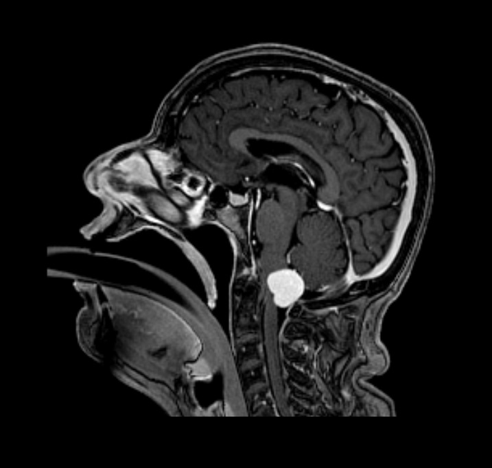
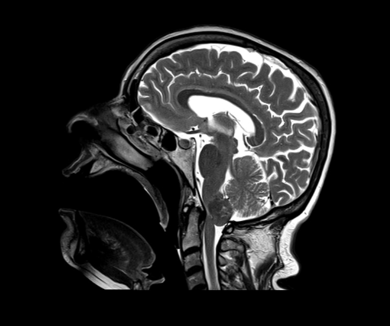
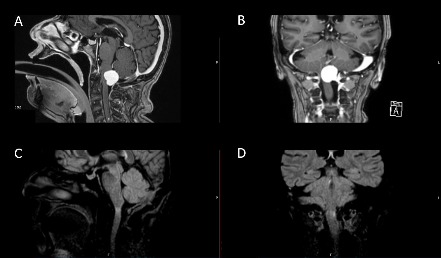
Results: A 73-year-old woman with hypertension and cervical dystonic tremor diagnosed ten years before was admitted to the hospital with one month history of occipital headache, progressive weakness in the left arm and gait imbalance. Neurological examination disclosed vertical-horizontal gaze-evoked nystagmus; monoparesis of the left superior limb grade 4 (MRC) with generalized hyperreflexia; dystonic posture of the left hand with slow distal sinuous movements; negative head tremor with cervical dystonia; left dysmetria in finger to nose test and unsteady gait with left lateropulsion. Brain and spinal MRI showed an expansive lesion located on the foramen magnum with homogeneous contrast enhancement causing marked compression of the bulbomedullary transition (image 1 and 2). The patient underwent surgery resection of the lesion which was confirmed to be a solitary fibrous tumor grade 1 (WHO – Classification of Tumors of the Central Nervous System). In the early post-operative period there was an improvement in neurological signs with persisting segmentary dystonia. The patient will maintain follow-up in the outpatient clinic.
Conclusion: To our knowledge this is the first case of a solitary fibrous tumor presenting with hand dystonia. We can discuss if this is a secondary dystonia or an anticipation phenomenon in a patient with primary dystonia.
To cite this abstract in AMA style:
S. Lopes, M. Santos, R. Pereira, M. Cambango, L. Oliveira, S. Varanda. Space-occupying lesion presenting with hand dystonia [abstract]. Mov Disord. 2023; 38 (suppl 1). https://www.mdsabstracts.org/abstract/space-occupying-lesion-presenting-with-hand-dystonia/. Accessed March 13, 2026.« Back to 2023 International Congress
MDS Abstracts - https://www.mdsabstracts.org/abstract/space-occupying-lesion-presenting-with-hand-dystonia/