Session Information
Date: Wednesday, September 25, 2019
Session Title: Neuroimaging
Session Time: 1:15pm-2:45pm
Location: Les Muses Terrace, Level 3
Objective: To evaluate impairments in the visual subnetwork in Parkinson’s disease (PD) patients with psychosis using BOLD fMRI
Background: Psychosis occurs in 20-70% of patients with PD and is commonly associated with exposure to dopamine agonists. Very few imaging studies have explored the basis for psychosis in PD and have usually included small sample sizes. In this work, we apply BOLD fMRI and a graph theory approach in a large cohort of age and gender-matched healthy controls (HC), PD patients without psychosis (PDNP), and PD patients with psychosis i.e. visual hallucinations (PDP).
Method: Forty-five HC (age=55.6±4.9 years, and levodopa daily dose, LEDD, = 0), 51 PDNP (age=57.9±7.0 years, LEDD= 569± 232 mg/d) and 47 PDP subjects (age=58.5± 8.2 years, LEDD= 678±300 mg/d) were assessed. In addition to the T1 data, fMRI BOLD acquisition parameters were: in-plane resolution = 1.65 mm , matrix size = 144 x 144, slices = 45, thickness = 3 mm, TR/TE = 2000/35 ms, 140 dynamics. All analysis was performed using Conn v18. Regions of the visual network were identified from Tunc et al. Local efficiency and clustering coefficient were calculated and compared per group at a cost = 0.15. All group comparisons were adjusted for age, gender, and LEDD. Significance at p < 0.05 was used on a two-sided t-test.
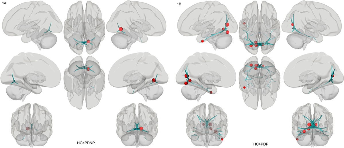
Results: Local efficiency of the visual network was significantly different between HC and PDNP (p = 0.01) (Figure 1A). The calcarine region (left) had significantly lower clustering coefficient in the PDNP group (0.74±0.20) compared to the HC group (0.83±0.17, p = 0.0005). There was no significant difference between the visual subnetwork in the PDNP and PDP groups. There was a significant difference between the HC and PDP group (p = 0.03) with lower clustering coefficients in the PDP group in multiple regions compared to HC (Figure 1B). The clustering coefficients in the left and right calcarine regions, left fusiform gyrus, left lingual gyrus, and left inferior temporal regions in the HC group were 0.83±0.17, 0.82±0.17, 0.69±0.21, 0.67±0.16, and 0.43±0.36. These values in the PDP group were 0.74±0.19, 0.71±0.17, 0.66±0.22, 0.64±0.23, and 0.39±0.32 respectively.
Conclusion: PD patients with psychosis have significantly altered connectivity in the visual subnetwork especially in the fusiform, lingual, and inferior temporal regions.
References: 1. Tunç, Birkan, et al. Philosophical Transactions of the Royal Society B: Biological Sciences 371.1688 (2016): 20150111. 2. Whitfield-Gabrieli, Susan, and Alfonso Nieto-Castanon. Brain connectivity 2.3 (2012): 125-141.
To cite this abstract in AMA style:
S. Rane, A. Lenka, S. Prasad, J. Saini, M. Ingalhalikar, P. Pal. Visual subnetwork dysfunction in Parkinson’s patients with primary visual hallucinations [abstract]. Mov Disord. 2019; 34 (suppl 2). https://www.mdsabstracts.org/abstract/visual-subnetwork-dysfunction-in-parkinsons-patients-with-primary-visual-hallucinations/. Accessed March 22, 2026.« Back to 2019 International Congress
MDS Abstracts - https://www.mdsabstracts.org/abstract/visual-subnetwork-dysfunction-in-parkinsons-patients-with-primary-visual-hallucinations/