Session Information
Date: Wednesday, June 22, 2016
Session Title: Neuroimaging (non-PD)
Session Time: 12:00pm-1:30pm
Location: Exhibit Hall located in Hall B, Level 2
Objective: To investigate longitudinal changes in clinical symptoms, 7T MRI and transcranial sonography (TCS) during chelating treatment in a Wilson disease (WD) patient.
Background: Pathophysiological causations and predictive biomarkers of clinical worsening in WD patients on anti-copper treatment are unknown. One of possible contributing factors may be brain iron accumulation. Our goal was to examine longitudinal changes in T2* relaxation time and echogenicity of deep grey matter structures as measures of iron content in a WD patient.
Methods: We examined a de-novo WD patient at baseline and followed him periodically for 30 months on anti-copper treatment. Clinical severity was assessed using the Unified Wilson Disease rating scale (UWDRS). The 7T MRI imaging protocol included a 3D magnetization prepared rapid gradient echo (MPRAGE, TE=3 ms; TR=2300 ms) and 2D multi-echo gradient echo (GRE, 8 echoes 4.1-25.5 ms; TR=1820 ms) for T2* parametric mapping. R2* values were measured in the globus pallidus (GP), putamen (Put), caudate nucleus (NC) and thalamus (Thal). TCS was performed and echogenicity indices of lentiform nucleus (NL) and NC were calculated using a software for TCS digital image analysis.
Results: 27-year old man developed ataxia and action tremor in hands and was diagnosed with WD (p.H1069Q homozygous mutation) three months later. At baseline there was pronounced NL hyperechogenicity bilaterally; R2* value in the GP (130) was 1.3 times increased compared to four age matched controls (mean 99.9±(SD)9.8). After penicillamin treatment initiation (increased to 900mg) ataxia and tremor progressed and generalized dystonia along with vertical gaze palsy appeared. After the switch to trientine (increased to 900mg) and later to Zinc(150mg)+trientine(600mg) his symptoms gradually improved and UWDRS score dropped from 104 to 56. R2* value gradually increased, particularly in Put (red arrows) and Thal (green arrows) suggesting iron accumulation. This was associated with thalamic atrophy and cavitation on MP-RAGE MRI (green arrows). TCS did not show any changes in NL and NC echogenicity during treatment. 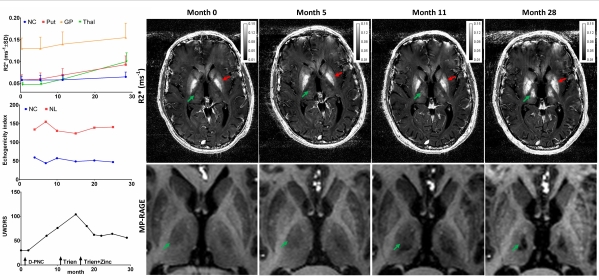
Conclusions: Our data suggest that increased brain iron concentration may be present already in de-novo WD while further iron deposition may accompany degenerative changes during unsuccessful anti-copper treatment. NL hyperechogenicity does not reflect R2* changes and may serve as a trait marker.
To cite this abstract in AMA style:
P. Dusek, D. Skoloudik, J. Maskova, T. Huelnhagen, R. Bruha, D. Zahorakova, T. Niendorf, E. Ruzicka, S.A. Schneider, J. Wuerfel. Iron accumulation in a Wilson disease patient: Longitudinal 7T MRI and transcranial sonography study [abstract]. Mov Disord. 2016; 31 (suppl 2). https://www.mdsabstracts.org/abstract/iron-accumulation-in-a-wilson-disease-patient-longitudinal-7t-mri-and-transcranial-sonography-study/. Accessed June 21, 2026.« Back to 2016 International Congress
MDS Abstracts - https://www.mdsabstracts.org/abstract/iron-accumulation-in-a-wilson-disease-patient-longitudinal-7t-mri-and-transcranial-sonography-study/