
Session Information
Date: Wednesday, June 22, 2016
Session Title: Neuroimaging (non-PD)
Session Time: 12:00pm-1:30pm
Location: Exhibit Hall located in Hall B, Level 2
Objective: We report a new case of isolated micrographia due to stroke involving the lenticular nucleus.
Background: Micrographia is a common sign in Parkinson´s disease while is a rare but recognised manifestation of an acute brain lesion commonly located in the left basal ganglia region.
Methods: Case report.
Results: We report a 57 year old right-handed woman with a permanent pacemaker because of an atrioventricular block who suddenly developed a right hemiplegia and aphasia. A Computed Tomography Angiography showed a thrombus occluding the left proximal middle cerebral artery (M1 segment). The patient received treatment with intravenous rtPa. Two hours later she was apparently asymptomatic. A computed tomography made 24 hours after treatment showed an infarct of the left lenticular nucleus and the head and body of the left caudate nucleus.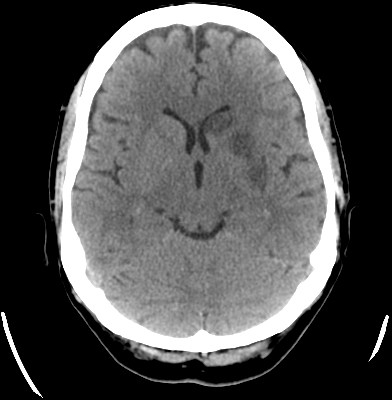 After going home and start their daily activity she realized that she had significant difficulties in writing with micrographia in which handwriting was microscopic from the outset.
 In the clinical examination just a slight impairment of rapid alternating movement without a clear decrease of amplitude of the right hand and fingers was observed. A DatScan was made, showing a complete absence of signal in the left striatal nuclei. A treatment with 300 mg per day of levodopa was not effective.
Conclusions: This case supports the evidence for the relationship between acute micrographia and focal brain lesion of the left basal ganglia region. It is important for clinicans to know that micrographia can represent an uncommon symptom of stroke.
To cite this abstract in AMA style:
P. Montero Escribano, M.J. Catalán Alonso, F. Alonso French, E. López Valdés, R. García Ramos, I. Parees Moreno. Isolated micrographia following stroke [abstract]. Mov Disord. 2016; 31 (suppl 2). https://www.mdsabstracts.org/abstract/isolated-micrographia-following-stroke/. Accessed November 15, 2026.« Back to 2016 International Congress
MDS Abstracts - https://www.mdsabstracts.org/abstract/isolated-micrographia-following-stroke/