Session Information
Date: Saturday, October 6, 2018
Session Title: Parkinson’s Disease: Clinical Trials, Pharmacology And Treatment
Session Time: 1:45pm-3:15pm
Location: Hall 3FG
Objective: To identify mitochondrial morphometric phenotypes in fibroblasts derived from patients with idiopathic Parkinson’s disease (IPD).
Background: Mitochondrial dysfunction (MD) has been proposed as cellular phenotype in patients with idiopathic Parkinson’s disease (IPD). However, study results remain controversial for fibroblasts derived from IPD. Here we aim to identify mitochondrial morphometric changes that eventually can be used in cell based drug screening assays targeting IPD.
Methods: Axilla skin punch biopsy derived fibroblasts from 41 IPD patients with a mean disease duration of 6.5 years (st.dev. 5.5 years) and from 21 healthy age-matched controls (HC) were stained with Hoechst, CellMask, and TMRM. Mitochondrial morphometrics were analyzed via automated confocal microscopy and in house developed computational image analysis (figure).
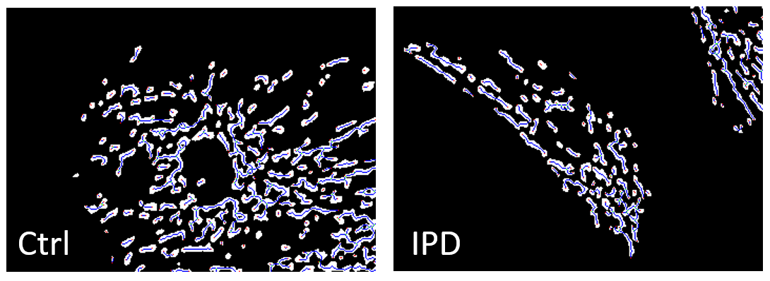
Results: Significant morphometric changes were seen in IPD fibroblasts. The number of mitochondria was increased (p=0.002) whereas the mitochondrial morphometric complexity was substantially decreased as visualized by reduction of mitochondrial perimeter (p=0.029) and mitochondrial form factor (p=0.013). Further analysis with established algorithms from computer vision and graph theory confirmed mitochondrial fragmentation: The mitochondrial skeleton corresponding to the final chain of pixels remaining after step by step erosion of the mitochondrial mask was significantly reduced in IPD fibroblasts (p=0.020). The number of mitochondrial nodes in the sense of graph theory applied to the mitochondrial skeleton reflects reduced mitochondrial branching in IPD (p=0.013). Finally, the mitochondrial node degree representing the average branch length in the mitochondrial skeleton network was also reduced (p<0.001). [figure1] Figure: (white) mitochondrial shapes, (blue) skeleton, (green) branch points, (red) endpoints. Note the reduced branching in the IPD patient sample.
Conclusions: Morphometric changes of IPD mitochondria with increased number of mitochondria on one side and reduced branching on the other side reflect mitochondrial fragmentation. This may be due to impaired mitophagy and/or also – at least temporary efficient – compensatory mechanisms, as also indicated by increased mitochondrial membrane potential. The results are in agreement with similar changes observed in submucosal enteric ganglia neurons of IPD patients (doi:10.1038/srep33117).
To cite this abstract in AMA style:
P. Antony, O. Boyd, K. Mommaerts, K. Sokolowska, M. Ostaszewski, A. Baumuratov, L. Longhino, F. Poulain, R. Krueger, R. Balling, N. Diederich. Mitochondrial morphometrics in idiopathic Parkinson‘s disease fibroblasts [abstract]. Mov Disord. 2018; 33 (suppl 2). https://www.mdsabstracts.org/abstract/mitochondrial-morphometrics-in-idiopathic-parkinsons-disease-fibroblasts/. Accessed July 23, 2026.« Back to 2018 International Congress
MDS Abstracts - https://www.mdsabstracts.org/abstract/mitochondrial-morphometrics-in-idiopathic-parkinsons-disease-fibroblasts/