Category: Dystonia: Pathophysiology, Imaging
Objective: To use task-based functional magnetic resonance imaging [fMRI] to examine body region-specific sensorimotor changes with the long-term goal of identifying potential unifying pathophysiological fMRI features of task-specific-focal-dystonia [TSFD] in our novel dataset of matched focal-hand-dystonia [FHD], laryngeal-dystonia [LD], and control [CTL] subjects. We hypothesize there will be brain activation differences between TSFD subtypes and CTL in areas related to voluntary motor control and sensorimotor integration, regardless of the task.
Background: TSFD is characterized by involuntary, localized muscle contractions during specific motor activities such as writing in FHD and speaking in LD [1]. It is unclear if a shared central mechanism contributes to different body regions being affected in subtypes of TSFD. Previous work using task-based fMRI has indicated abnormal activation in body-region-specific sensorimotor networks separately in FHD and LD against CTL [2-5]. However, the relationship between TSFD subgroups has not been well-described.
Method: Nineteen adults with LD (59.55 ± 13.47 years, 8 female), 14 with FHD (55.43 ± 15.82, 6 female), and 20 CTL (53.5 ± 13.95 years, 4 females) completed fMRI tasks in a Siemens 3T scanner. Brain activation was measured using blood-oxygen-level-dependent responses to phonation, left-finger tapping, and right-finger-tapping. Brain images for four FHD subjects were mirrored, so the right-hand always represents the affected hand.
Results: Results from this preliminary analysis indicate that compared to CTL, TSFD demonstrated increased activation in associative cortical areas during dystonia-related task performance. During phonation, the LD > CTL contrast demonstrated increased activation in the posterior parietal cortex (inferior parietal lobule [IPL] encompassing the supramarginal and angular gyrus, left superior lobule). During finger-tapping, the FHD > CTL contrast also demonstrated increased activation in the IPL and the superior and middle frontal gyri. Notably, this increased activity was only observed in the ipsilateral hemisphere for affected-side finger-tapping in FHD [Fig1].
Conclusion: These findings underscore the involvement of parietal sensory association areas during dystonia-related tasks and suggest that sensory processing abnormalities may contribute to or be associated with TSFD. Further results comparing TSFD subtypes will be presented.
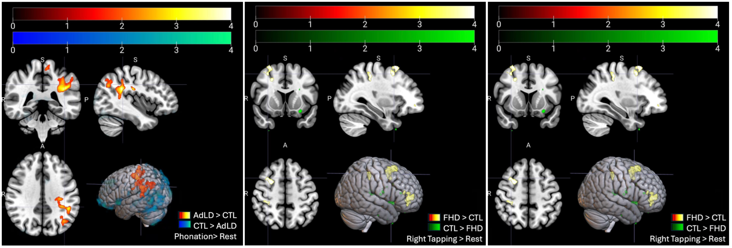
Fig1
References: 1. Albanese A, Bhatia K, Bressman SB, et al. Phenomenology and classification of dystonia: a consensus update. Mov Disord Off J Mov Disord Soc. 2013;28(7):863-873. doi:10.1002/mds.25475
2. Pujol J, Roset-Llobet J, Rosinés-Cubells D, et al. Brain cortical activation during guitar-induced hand dystonia studied by functional MRI. NeuroImage. 2000;12(3):257-267. doi:10.1006/nimg.2000.0615
3. Preibisch C, Berg D, Hofmann E, Solymosi L, Naumann M. Cerebral activation patterns in patients with writer’s cramp: a functional magnetic resonance imaging study. J Neurol. 2001;248(1):10-17. doi:10.1007/s004150170263
4. Delnooz CCS, Helmich RC, Medendorp WP, Van de Warrenburg BPC, Toni I. Writer’s cramp: increased dorsal premotor activity during intended writing. Hum Brain Mapp. 2013;34(3):613-625. doi:10.1002/hbm.21464
5. Simonyan K, Ludlow CL. Abnormal Activation of the Primary Somatosensory Cortex in Spasmodic Dysphonia: An fMRI Study. Cereb Cortex N Y NY. 2010;20(11):2749-2759. doi:10.1093/cercor/bhq023
To cite this abstract in AMA style:
B. Huynh, Y. Wang, M. Chen, E. Narinsky, YL. Kuo, T. Kimberley. Task-Based Functional Imaging in Task-Specific Focal Dystonia during Dystonia-Related and -Unrelated Tasks [abstract]. Mov Disord. 2024; 39 (suppl 1). https://www.mdsabstracts.org/abstract/task-based-functional-imaging-in-task-specific-focal-dystonia-during-dystonia-related-and-unrelated-tasks/. Accessed May 26, 2026.« Back to 2024 International Congress
MDS Abstracts - https://www.mdsabstracts.org/abstract/task-based-functional-imaging-in-task-specific-focal-dystonia-during-dystonia-related-and-unrelated-tasks/