Category: MSA, PSP, CBS: Neuroimaging
Objective: This study aims to explore possible glymphatic dysfunction using diffusion tensor image analysis along the perivascular space (DTI-ALPS) in patients with progressive supranuclear palsy (PSP) and their relationship with clinical outcomes.
Background: PSP is an atypical parkinsonian syndrome classified as a 4-repeat-tauopathy [1]. The DTI-ALPS indices have been proposed as a non-invasive local proxy of the glymphatic system activity [2], which is well-known as a waste drainage system of interstitial solutes like tau neurofibrillary tangles [3]. Nevertheless, this measure and its relationship with clinical features have been barely explored in PSP.
Method: Nineteen patients with PSP and 31 sex-matched healthy controls (HC) were assessed using diffusion-weighted magnetic resonance imaging (MRI). The severity and progression of PSP were clinically evaluated using the Progressive Supranuclear Palsy Rating Scale (PSPRS). Automated DTI-ALPS indices were computed for the left and right hemispheres, and bilaterally. Between-group comparisons were performed for the demographic, clinical and DTI-ALPS variables, controlling by age and years of education. Correlations between demographic and clinical features and DTI-ALPS indices were computed. All the results were corrected by false discovery rate (FDR, p<0.05).
Results: Patients with PSP showed significantly decreased DTI-ALPS indices bilaterally (meanHC=1.68±0.21, meanPSP=1.44±0.12, F=-3.284, pFDR=0.005, d=1.416), and in the right (meanHC=1.66±0.22, meanPSP=1.42±0.13, F=-2.937, pFDR=0.005, d=1.309) and left (meanHC=1.71±0.24, meanPSP=1.45±0.17, F=-2.985, pFDR=0.005, d=1.233) hemispheres after controlling by age and education [Figure1]. Clinically, patients with PSP showed higher non-motor symptoms, and more neuropsychiatric comorbidities, apathy and depression than HC [Table1]. The DTI-ALPS index in the left hemisphere was negatively correlated with the PSPRS (r=-0.604, pFDR=0.041) controlled by age and education [Figure2A], and the DTI-ALPS index in the right hemisphere negatively correlated with age (r=-0.509, p=0.026) in patients with PSP [Figure2B]. No other correlations were found in the HC or PSP group.
Conclusion: The current results suggest a dysfunctional perivascular glymphatic activity, as decreased DTI-ALPS indices, in patients with PSP, which was associated with age and a clinical measure of severity and progression of the disease.
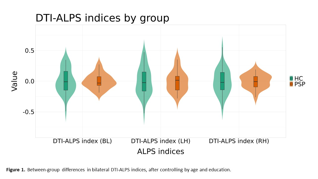
Figure 1
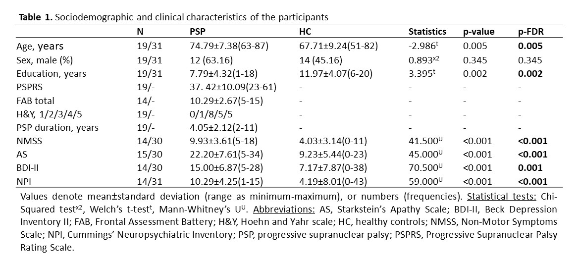
Table 1
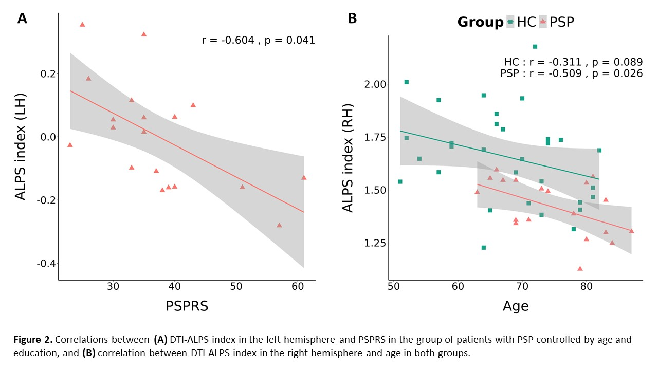
Figure 2
References: [1] Williams DR, Lees AJ. Progressive supranuclear palsy: clinicopathological concepts and diagnostic challenges. Vol. 8, The Lancet Neurology. 2009. p. 270–9.
[2] Taoka T, Masutani Y, Kawai H, Nakane T, Matsuoka K, Yasuno F, et al. Evaluation of glymphatic system activity with the diffusion MR technique: diffusion tensor image analysis along the perivascular space (DTI-ALPS) in Alzheimer’s disease cases. Jpn J Radiol. 2017 Apr 1;35(4):172–8.
[3] Ota M, Sato N, Takahashi Y, Shigemoto Y, Kimura Y, Nakaya M, et al. Correlation between the Regional Brain Volume and Glymphatic System Activity in Progressive Supranuclear Palsy. Dement Geriatr Cogn Disord. 2023 Jul 1;52(3):177–83.
To cite this abstract in AMA style:
J. Pardo, C. Martín-Barceló, C. Falcon, R. Sala-Llonch, I. Roura, A. Campabadal, N. Bargalló, C. Painous, M. Fernández, O. Brengaret, C. Brenlla, A. Cámara, E. Muñoz, F. Valldeoriola, MJ. Martí, Y. Compta, C. Junqué, B. Segura. Association between Imaging Glymphatic Dysfunction Markers and Clinical Symptoms in Progressive Supranuclear Palsy [abstract]. Mov Disord. 2025; 40 (suppl 1). https://www.mdsabstracts.org/abstract/association-between-imaging-glymphatic-dysfunction-markers-and-clinical-symptoms-in-progressive-supranuclear-palsy/. Accessed July 7, 2026.« Back to 2025 International Congress
MDS Abstracts - https://www.mdsabstracts.org/abstract/association-between-imaging-glymphatic-dysfunction-markers-and-clinical-symptoms-in-progressive-supranuclear-palsy/