Category: Ataxia
Objective: The aim of this study was to evaluate clinical manifestations and magnetic resonance imaging (MRI) changes in patients with ANO10 gene mutations.
Background: Neurodegenerative cerebellar ataxias are a heterogeneous group of disorders that predominantly affect the cerebellum. Biallelic variants in the ANO10 gene cause an autosomal recessive cerebellar ataxia designated ATX-ANO10 (OMIM #613728) and only 94 cases have been reported worldwide.
Method: We enrolled 7 patients with the biallelic c.1150_1151del variant in the ANO10 gene and matched for age and sex with 14 healthy controls (HC) who were clinically examined at the Neurology Clinic, University Clinical Centre of Serbia. The neurological examination included SARA, INAS, ACE-R, MMSE, HARS and HDRS scales. All participants underwent MRI of the brain on a 1.5T MRI scanner (Philips Medical Systems, Achieva). Unconventional MRI methods such as voxel-based morphometry (VBM) and spatially unbiased infratentorial template (SUIT, VBM) were used to analyse the morphological and topographical distribution of grey matter (GM) and white matter (WM) changes. Brainstem and superior cerebellar peduncles (SCP) volumes were estimated and compared using Freesurfer. MRI results were obtained by researchers from San Raffaele, University of Milan, Italy.
Results: The mean age at onset of the patients with ANO10 mutation was 16.3±5.3 years, while duration of the disease was 25±12.3 years. All patients presented with cerebellar syndrome, pyramidal signs and ocumolotor abnormalities. The results of the different scales used are shown in Table 1. VBM and SUIT VBM of GM and WM showed that participants with ANO10 mutation had a significant difference compared to HC (p <0.05, ANCOVA, age and sex as covariates) in isolated brainstem volumes (medulla, pons, midbrain, SCP), as well as total brainstem volume. Patients with mutation in ANO10 gene also had involvement of supratentorial regions: right inferior orbitofrontal region, insula, operculum, left supplementary motor area, right and left medial frontal regions, temporal regions.
Conclusion: Our study has shown that ANO10 mutation carriers have prominent cerebellar and brainstem atrophy, as well as involvement of the supratentorial regions, which may be associated with clinical manifestations beyond cerebellar involvement.
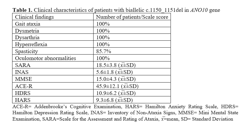
Table 1.
To cite this abstract in AMA style:
A. Milovanović, O. Tamaš, N. Mazalica, S. Pisano, S. Basaia, F. Agosta, M. Filippi, V. Kostić, N. Dragašević-Mišković. Clinical and Neuroimaging Findings in ANO10-Related Cerebellar Ataxia: Evidence of Widespread Brain Atrophy [abstract]. Mov Disord. 2025; 40 (suppl 1). https://www.mdsabstracts.org/abstract/clinical-and-neuroimaging-findings-in-ano10-related-cerebellar-ataxia-evidence-of-widespread-brain-atrophy/. Accessed July 10, 2026.« Back to 2025 International Congress
MDS Abstracts - https://www.mdsabstracts.org/abstract/clinical-and-neuroimaging-findings-in-ano10-related-cerebellar-ataxia-evidence-of-widespread-brain-atrophy/