Category: Ataxia
Objective: To characterize the structural brain signature in SCA6 patients.
Background: Spinocerebellar ataxias (SCA) refer to a group of autosomal dominant ataxic disorders that result from the degeneration of the cerebellum and its connections (1). SCA6 is described as the prototype of pure cerebellar ataxia, with preservation of other brain regions (2). However, the calcium receptor subunit affected in SCA6 appears to be ubiquitous in neurons throughout the brain (3). Additionally, there are observations of clinical involvement of non-cerebellar systems, structural cerebral damage and hypometabolism in various brain areas (4-6).
Method: Eighteen SCA6 patients underwent cross-sectional analyses using multimodal MRI-based techniques, which combined cerebral and cerebellar volumetric analyses with diffusion tensor imaging (DTI). Furthermore, we investigated whether structural abnormalities correlated with clinical findings.
Results: Individuals with SCA6, compared to non-ataxic controls, exhibited significant volumetric reduction in cerebellar white matter, cortex, and several lobules. There was increased axial diffusivity (AD) in the left inferior cerebellar peduncle, left middle cerebellar peduncle, left superior cerebellar peduncle, left superior corona radiata, left fornix-stria terminalis, left sagittal stratum, left genu of the corpus callosum, left midbrain, right cerebral peduncle, right fornix-stria terminalis, right middle cerebellar peduncle, right body of the corpus callosum, right midbrain, and left optic tract [figure 1]. Significant correlations were found between AD and the Inventory of Non-ataxia Symptoms Count and the Epworth Sleepiness Scale [figure 2].
Conclusion: We provided valuable insights into the extracerebellar structural abnormalities associated with SCA6. DTI-based analyses of cerebellar connections and supratentorial structures emerge as potential sources of biomarkers for SCA6.
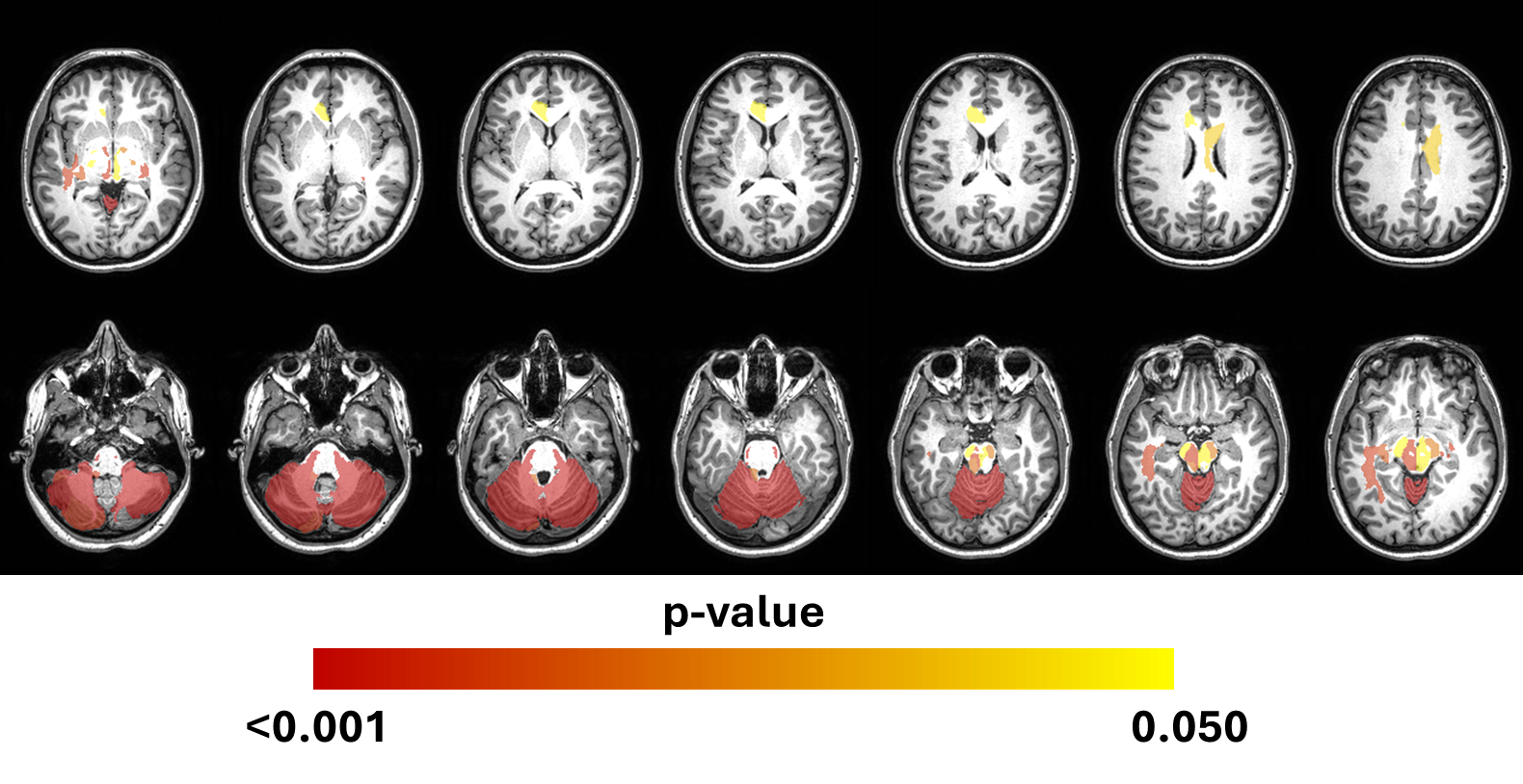
figure 1
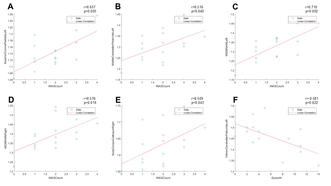
figure 2
References: 1. Schöls L, Bauer P, Schmidt T, Schulte T, Riess O. Autosomal dominant cerebellar ataxias: clinical features, genetics, and pathogenesis. Lancet Neurol. 2004 May;3(5):291–304.
2. Ishikawa K, Watanabe M, Yoshizawa K, Fujita T, Iwamoto H, Yoshizawa T, et al. Clinical, neuropathological, and molecular study in two families with spinocerebellar ataxia type 6 (SCA6). J Neurol Neurosurg Psychiatry. 1999 Jul;67(1):86–9.
3. Hillman D, Chen S, Aung TT, Cherksey B, Sugimori M, Llinás RR. Localization of P-type calcium channels in the central nervous system. Proc Natl Acad Sci U S A. 1991 Aug 15;88(16):7076–80.
4. Koeppen AH. The pathogenesis of spinocerebellar ataxia. Cerebellum. 2005;4(1):62–73.
5. Oh M, Kim JS, Oh JS, Lee CS, Chung SJ. Different subregional metabolism patterns in patients with cerebellar ataxia by 18F-fluorodeoxyglucose positron emission tomography. PLoS One. 2017;12(3):e0173275.
6. Schöls L, Krüger R, Amoiridis G, Przuntek H, Epplen JT, Riess O. Spinocerebellar ataxia type 6: genotype and phenotype in German kindreds. J Neurol Neurosurg Psychiatry. 1998 Jan;64(1):67–73.
To cite this abstract in AMA style:
B. Massuyama, T. Rezende, M. Junior, O. Barsottini, J. Pedroso. Brain Structural Impairment in Spinocerebellar Ataxia Type 6: Not Restricted to the Cerebellum [abstract]. Mov Disord. 2025; 40 (suppl 1). https://www.mdsabstracts.org/abstract/brain-structural-impairment-in-spinocerebellar-ataxia-type-6-not-restricted-to-the-cerebellum/. Accessed July 7, 2026.« Back to 2025 International Congress
MDS Abstracts - https://www.mdsabstracts.org/abstract/brain-structural-impairment-in-spinocerebellar-ataxia-type-6-not-restricted-to-the-cerebellum/