Objective: We recently showed that HMGB2 is secreted during ferroptosis and as ferroptosis is involved in the etiology of PD, we have examined the possibility that HMGB2 is secreted during PD so that it might serve as a new PD biomarker.
Background: PD is a progressive neurodegenerative disorder characterized by motor and non-motor symptoms, with early diagnosis remaining a challenge. The most widely used biomarker, phosphorylated alpha-synuclein, has 94% specificity. HMGB proteins, particularly HMGB1, have been linked to neuroinflammation, oxidative stress, and apoptosis—key processes in PD pathogenesis.
Method: HMGB2 peptides were analyzed using stable isotope dilution with immunoprecipitation (IP), trypsin digestion, and LC-parallel reaction monitoring (PRM)/MS. Experiments were conducted on a Thermo Scientific Q Exactive HF Orbitrap MS coupled to a Dionex Ultimate 3000 RSLCnano system. SILIB-HMGB2, used as the internal standard, was expressed in E. coli using [13C,15N]-labeled medium. Ferroptosis was induced in SH-SY5Y cells using rotenone (20 µM) or erastin (50 µM).
Results: Media was separated from the cells after incubations with the ferroptosis inducers rotenone (20 microM) or erastin (50 microM), SILIB internal standard added (50 ng), the media purified by IP, and digested with trypsin overnight at 37 oC. Standard curve samples for tryptic digests were prepared in media in the range 40 to 1200 ng using peptides H31PDASVNFSEFSK43 and S115EHPGLSIGDTAK127. Light to heavy peptide ratios were calculated for the sum of the three most intense PRM transitions for each peptide. Results show 3.68 ± 0.09 mg/106 cells (n = 3) and 3.87 ± 0.13 mg/106 cells (n = 3) for 24-h incubation with erastin or rotenone, respectively. Plasma samples from PD patients (n=5) had undetectable unmodified HMGB2 but contained mono-phosphorylated (MH2²⁺ = m/z 772.825) and bis-phosphorylated (MH2²⁺ = m/z 812.808) H31PDSSVNFAEFSK43 at S-35 or S-35 and S-42. Dephosphorylation with lambda protein phosphatase led to a substantial decrease in phosphorylated peptides and the appearance of unmodified H31PDSSVNFAEFSK43 (MH2²⁺ = m/z 732.841). Chymotryptic digestion confirmed phosphorylation at S-42 and S-35. Synthetic standards will be required for definitive site confirmation.
Conclusion: Phosphorylated HMGB2 has potential to be a diagnostic biomarker for PD and may help distinguish PD from other neurodegenerative disorders.
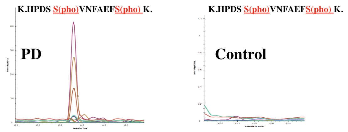
Phosphorylated HMGB protein in PD plasma samples
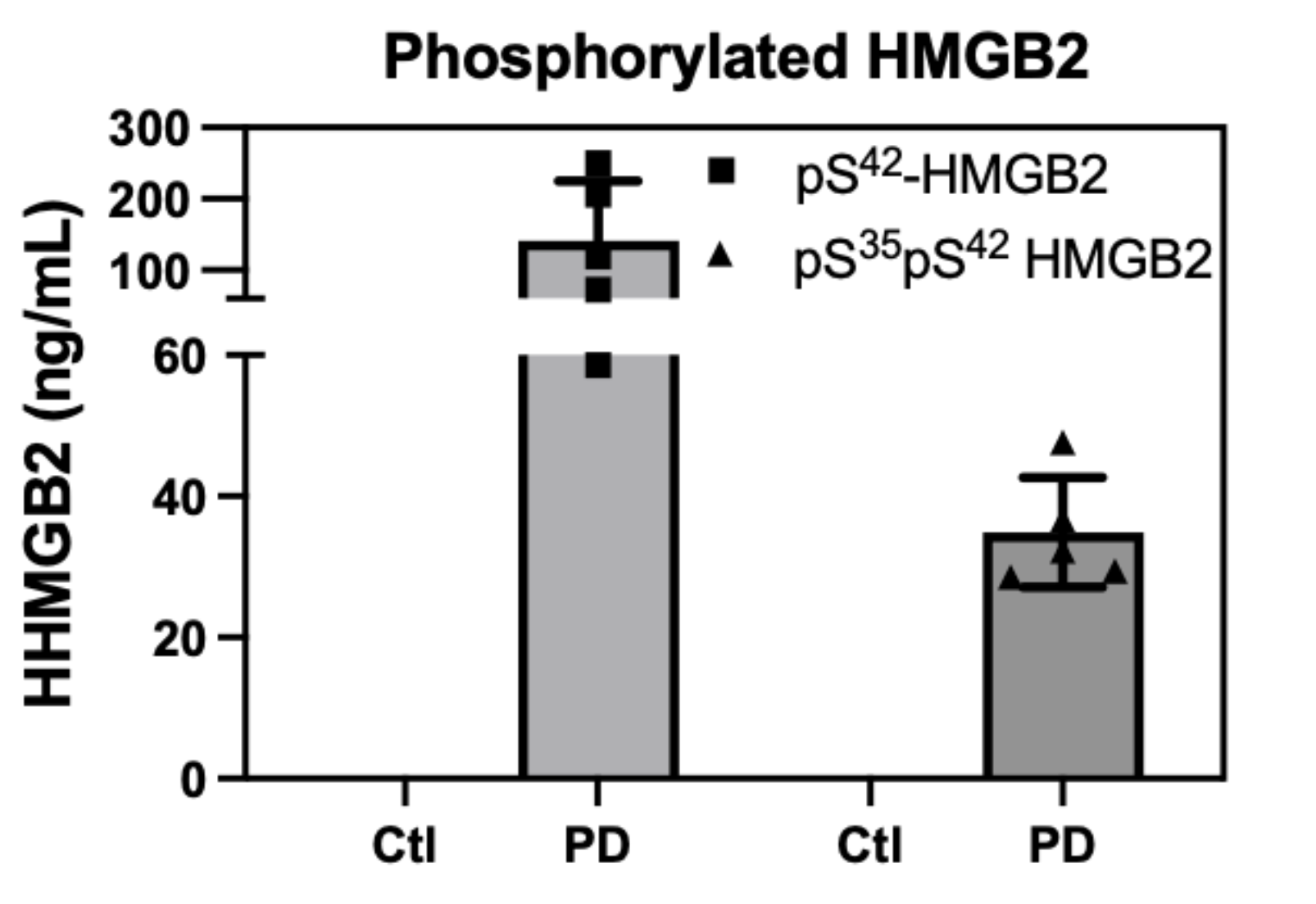
Phosphorylated HMGB2 in PD plasma (n=5)
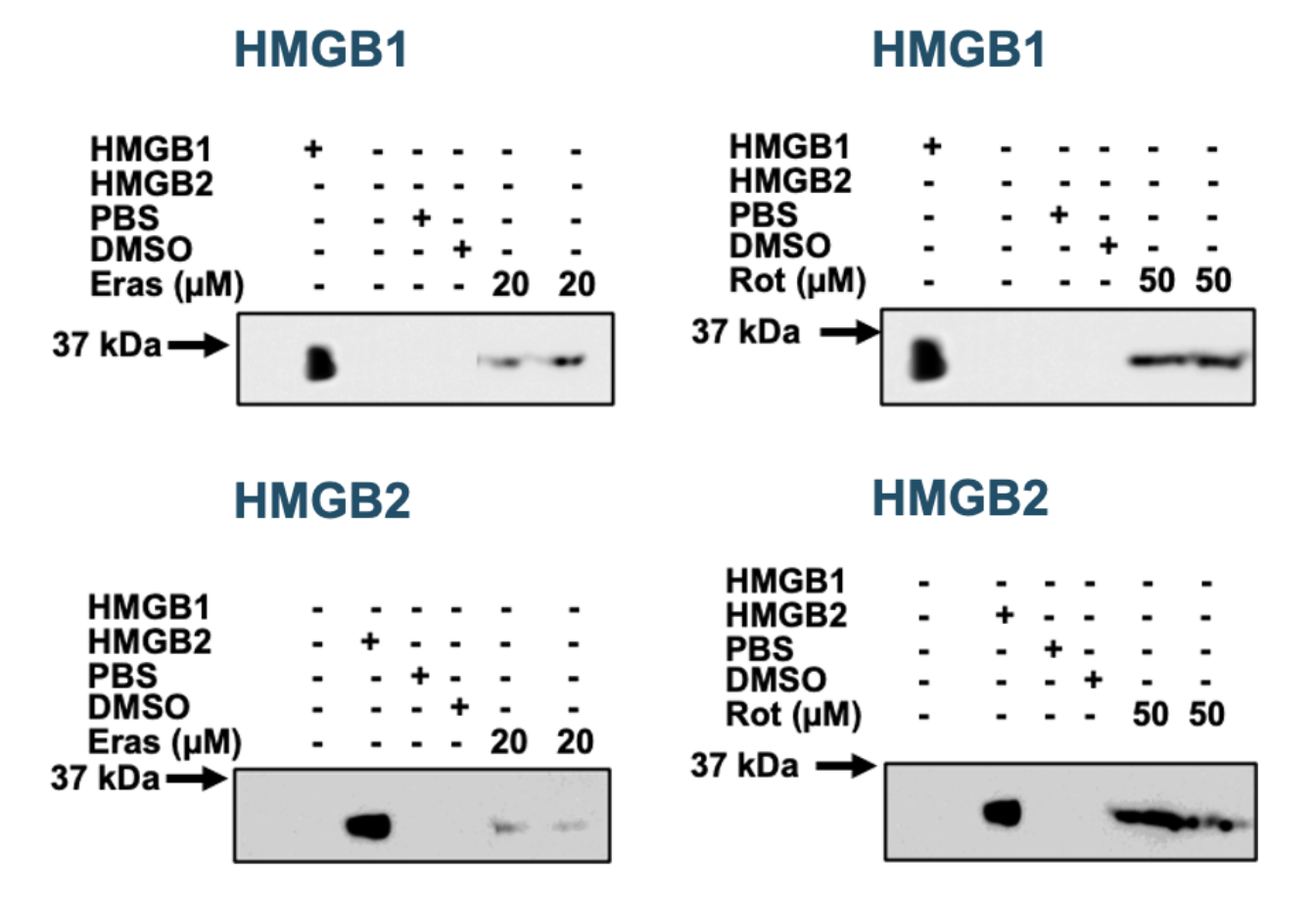
HMGBs secretion
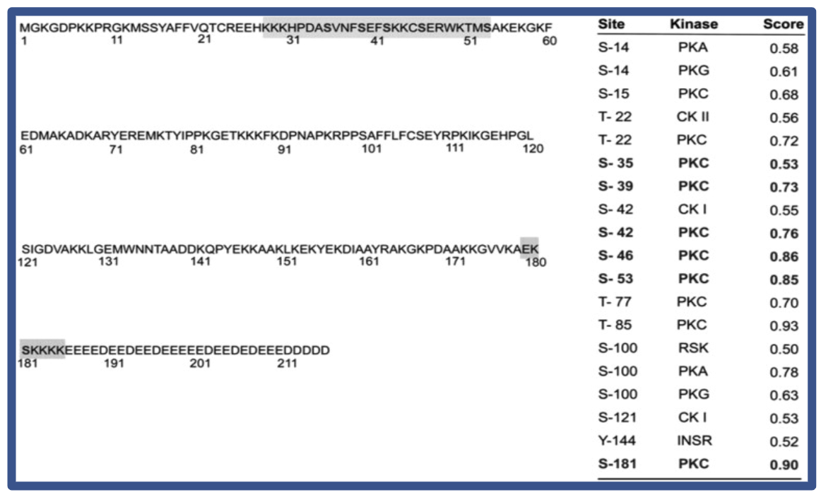
Prediction of phosphorylated sites in HMGB
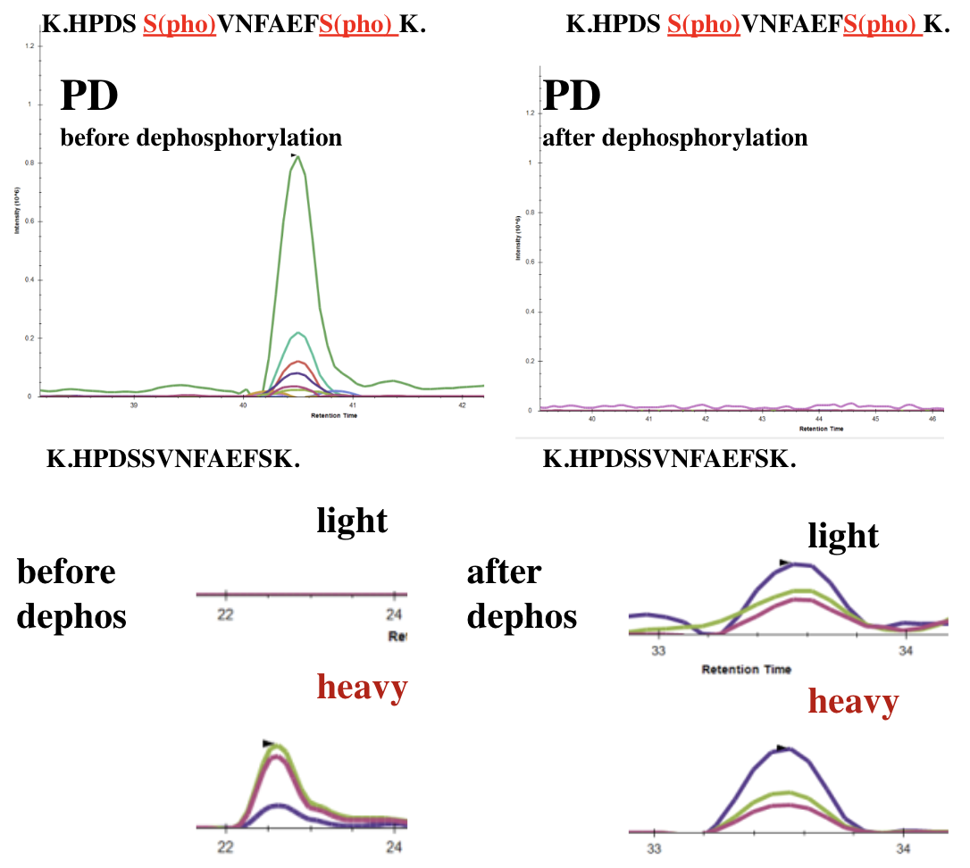
LC/MS signals before and after dephosphorylation
To cite this abstract in AMA style:
J. Fan, J. Liu, C. Mesaros, I. Blair. Phosphorylated High Mobility Group Box Protein 2 (HMGB2) as a Potential Biomarker for Parkinson’s Disease [abstract]. Mov Disord. 2025; 40 (suppl 1). https://www.mdsabstracts.org/abstract/phosphorylated-high-mobility-group-box-protein-2-hmgb2-as-a-potential-biomarker-for-parkinsons-disease/. Accessed July 10, 2026.« Back to 2025 International Congress
MDS Abstracts - https://www.mdsabstracts.org/abstract/phosphorylated-high-mobility-group-box-protein-2-hmgb2-as-a-potential-biomarker-for-parkinsons-disease/