Category: Parkinson's disease: Neuroimaging
Objective: This study investigates the effects of a novel TBS paradigm on large-scale brain network connectivity in PD patients.
Background: Parkinson’s disease (PD) is a neurodegenerative disorder characterized by progressive motor dysfunction and cognitive decline. While deep brain stimulation (DBS) of the subthalamic nucleus (STN) at gamma frequencies effectively manages motor symptoms, it has been linked to cognitive deficits. Theta-burst stimulation (TBS) has shown promise in enhancing cognitive function without impairing motor control. Functional magnetic resonance imaging (fMRI) is a powerful tool for assessment of brain network connectivity and has been used to characterize differences in DBS protocols.
Method: Eight PD patients [table1] with existing STN-DBS implants underwent resting-state fMRI under two stimulation conditions: (1) standard gamma-frequency and (2) theta-burst DBS [figure1]. Stimulation order was randomized, with a 10-minute washout period between conditions. We assessed connectivity within and between seven cortical networks, including the visual, somatomotor, dorsal attention (DAN), salience, limbic, control, and default mode networks (DMN).
Results: Overall network integrity was preserved in gamma and TBS stimulation paradigms, with stronger within-network connectivity compared to between-network connectivity [figure2]. However, TBS significantly increased anti-correlation between the DMN and the DAN (p = 0.0098, FDR-corrected) and between the DMN and the visual network (p =0.024, FDR-corrected), suggesting enhanced segregation of cognitive and attention networks. TBS increased connectivity between the DAN and salience network (p =0.024, FDR-corrected) [figure3].
Conclusion: Our study provides novel insights into the effects of TBS on large-scale brain network dynamics in PD patients undergoing DBS. The observed anti-correlation between the DMN and both the DAN and VIS aligns with evidence linking excessive DMN activity to cognitive interference and attentional deficits in PD, while increased DAN-SAL connectivity may help restore balance between attention and salience networks, which are often disrupted in PD and associated with cognitive rigidity and impaired motor function. These results highlight the potential of TBS as a neuromodulation approach for optimizing brain network organization in PD which may be further validated through motor and cognitive testing.
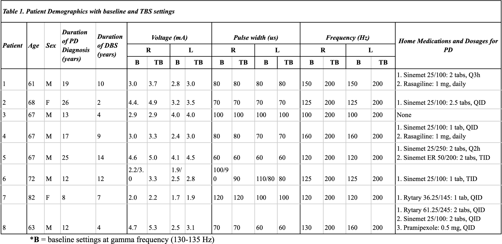
table1
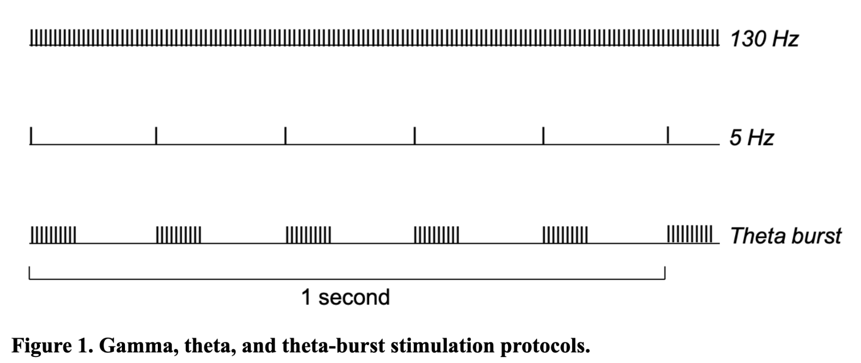
figure1
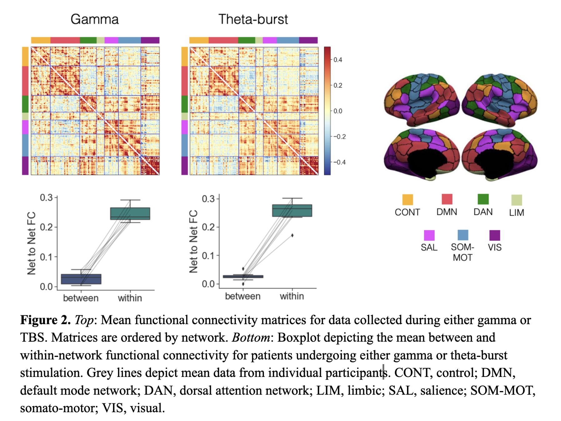
figure2
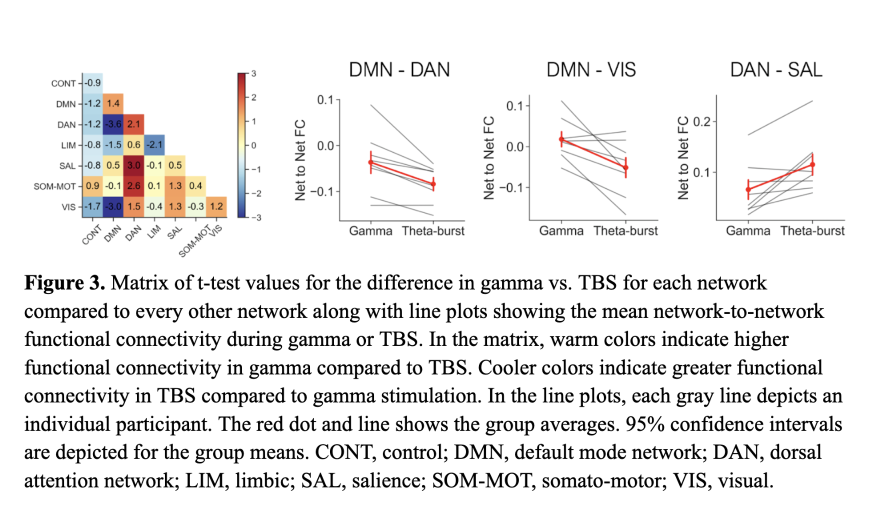
figure3
References: [1] Degirmenci Y, Angelopoulou E, Georgakopoulou VE, Bougea A. Cognitive impairment in Parkinson’s disease: An updated overview focusing on emerging pharmaceutical treatment approaches. Medicina (Kaunas). 2023;59. doi:10.3390/medicina59101756
[2] Tolosa E, Garrido A, Scholz SW, Poewe W. Challenges in the diagnosis of Parkinson’s disease. Lancet Neurol. 2021;20: 385–397.
[3] Horn MA, Gulberti A, Gülke E, Buhmann C, Gerloff C, Moll CKE, et al. A new stimulation mode for deep brain stimulation in Parkinson’s disease: Theta burst stimulation. Mov Disord. 2020;35: 1471–1475.
[4] Gülke E, Horn MA, Caffier J, Pinnschmidt H, Hamel W, Moll CKE, et al. Comparison of subthalamic unilateral and bilateral theta burst deep brain stimulation in Parkinson’s disease. Front Hum Neurosci. 2023;17: 1233565.
[5] Barbero JA, Unadkat P, Choi YY, Eidelberg D. Functional brain networks to evaluate treatment responses in Parkinson’s disease. Neurotherapeutics. 2023;20: 1653–1668.
[6] Schindlbeck KA, Lucas-Jiménez O, Tang CC, Morbelli S, Arnaldi D, Pardini M, et al. Metabolic network abnormalities in drug‐naïve Parkinson’s disease. Mov Disord. 2020;35: 587–594.
[7] Horn A, Wenzel G, Irmen F, Huebl J, Li N, Neumann W-J, et al. Deep brain stimulation induced normalization of the human functional connectome in Parkinson’s disease. Brain. 2019;142: 3129–3143.
[8] Blonder LX, Slevin JT. Emotional dysfunction in Parkinson’s disease. Behav Neurol. 2011;24: 201–217.
[9] Kahan J, Urner M, Moran R, Flandin G, Marreiros A, Mancini L, et al. Resting state functional MRI in Parkinson’s disease: the impact of deep brain stimulation on “effective” connectivity. Brain. 2014;137: 1130–1144.
[10] Lai H-Y, Younce JR, Albaugh DL, Kao Y-CJ, Shih Y-YI. Functional MRI reveals frequency-dependent responses during deep brain stimulation at the subthalamic nucleus or internal globus pallidus. Neuroimage. 2014;84: 11–18.
[11] Boutet A, Madhavan R, Elias GJB, Joel SE, Gramer R, Ranjan M, et al. Predicting optimal deep brain stimulation parameters for Parkinson’s disease using functional MRI and machine learning. Nat Commun. 2021;12: 3043.
[12] Weil RS, Schrag AE, Warren JD, Crutch SJ, Lees AJ, Morris HR. Visual dysfunction in Parkinson’s disease. Brain. 2016;139: 2827–2843.
[13] Sheng K, Fang W, Su M, Li R, Zou D, Han Y, et al. Altered spontaneous brain activity in patients with Parkinson’s disease accompanied by depressive symptoms, as revealed by regional homogeneity and functional connectivity in the prefrontal-limbic system. PLoS One. 2014;9: e84705.
[14] Ragothaman A, Mancini M, Nutt JG, Wang J, Fair DA, Horak FB, et al. Motor networks, but also non-motor networks predict motor signs in Parkinson’s disease. NeuroImage Clin. 2023;40: 103541.
[15] Boon LI, Hepp DH, Douw L, van Geenen N, Broeders TAA, Geurts JJG, et al. Functional connectivity between resting-state networks reflects decline in executive function in Parkinson’s disease: A longitudinal fMRI study. NeuroImage Clin. 2020;28: 102468.
[16] Borst JP, Anderson JR. Using model-based functional MRI to locate working memory updates and declarative memory retrievals in the fronto-parietal network. Proc Natl Acad Sci U S A. 2013;110: 1628–1633.
[17] Vincent JL, Kahn I, Snyder AZ, Raichle ME, Buckner RL. Evidence for a frontoparietal control system revealed by intrinsic functional connectivity. J Neurophysiol. 2008;100: 3328–3342.
[18] Cascone AD, Langella S, Sklerov M, Dayan E. Frontoparietal network resilience is associated with protection against cognitive decline in Parkinson’s disease. Commun Biol. 2021;4: 1021.
[19] Schaefer A, Kong R, Gordon EM, Laumann TO, Zuo X-N, Holmes AJ, et al. Local-global parcellation of the human cerebral cortex from intrinsic functional connectivity MRI. Cereb Cortex. 2018;28: 3095–3114.
[20] Lam J, Lee J, Williams M, Cohn M, Wilson M, Mark C, et al. Cognitive effects of theta frequency bilateral subthalamic nucleus stimulation in Parkinson’s disease: A pilot study. Brain Stimul. 2021;14: 230–240.
[21] Fiorenzato E, Strafella AP, Kim J, Schifano R, Weis L, Antonini A, et al. Dynamic functional connectivity changes associated with dementia in Parkinson’s disease. Brain. 2019;142: 2860–2872.
[22] Díez-Cirarda M, Strafella AP, Kim J, Peña J, Ojeda N, Cabrera-Zubizarreta A, et al. Dynamic functional connectivity in Parkinson’s disease patients with mild cognitive impairment and normal cognition. NeuroImage Clin. 2018;17: 847–855.
[23] Baggio H-C, Sala-Llonch R, Segura B, Marti M-J, Valldeoriola F, Compta Y, et al. Functional brain networks and cognitive deficits in Parkinson’s disease. Hum Brain Mapp. 2014;35: 4620–4634.
[24] Bai Y, Diao Y, Gan L, Zhuo Z, Yin Z, Hu T, et al. Deep brain stimulation modulates multiple abnormal resting-state network connectivity in patients with Parkinson’s disease. Front Aging Neurosci. 2022;14: 794987.
[25] Anticevic A, Cole MW, Murray JD, Corlett PR, Wang X-J, Krystal JH. The role of default network deactivation in cognition and disease. Trends Cogn Sci. 2012;16: 584–592.
[26] Shang Y, Chang D, Zhang J, Peng W, Song D, Gao X, et al. Theta-burst transcranial magnetic stimulation induced functional connectivity changes between dorsolateral prefrontal cortex and default-mode-network. Brain Imaging Behav. 2020;14: 1955–1963.
[27] Zhang Z, Zheng H, Liang K, Wang H, Kong S, Hu J, et al. Functional degeneration in dorsal and ventral attention systems in amnestic mild cognitive impairment and Alzheimer’s disease: an fMRI study. Neurosci Lett. 2015;585: 160–165.
[28] Lee DJ, Drummond NM, Saha U, De Vloo P, Dallapiazza RF, Gramer R, et al. Acute low frequency dorsal subthalamic nucleus stimulation improves verbal fluency in Parkinson’s disease. Brain Stimul. 2021;14: 754–760.
[29] Shang S ’an, Wang L, Xu Y, Zhang H, Chen L, Dou W, et al. Optimization of structural connectomes and scaled patterns of structural-functional decoupling in Parkinson’s disease. Neuroimage. 2023;284: 120450.
[30] Baik K, Kim YJ, Park M, Chung SJ, Sohn YH, Jeong Y, et al. Functional brain networks of minor and well-structured major hallucinations in Parkinson’s disease. Mov Disord. 2024;39: 318–327.
[31] Bhome R, Thomas GEC, Zarkali A, Weil RS. Structural and functional imaging correlates of visual hallucinations in Parkinson’s disease. Curr Neurol Neurosci Rep. 2023;23: 287–299.
[32] Chai XJ, Ofen N, Gabrieli JDE, Whitfield-Gabrieli S. Selective development of anticorrelated networks in the intrinsic functional organization of the human brain. J Cogn Neurosci. 2014;26: 501–513.
[33] Cohen JR, D’Esposito M. The segregation and integration of distinct brain networks and their relationship to cognition. J Neurosci. 2016;36: 12083–12094.
[34] Rahnev D, Kok P, Munneke M, Bahdo L, de Lange FP, Lau H. Continuous theta burst transcranial magnetic stimulation reduces resting state connectivity between visual areas. J Neurophysiol. 2013;110: 1811–1821.
[35] Cohan R, Rafique SA, Stoby KS, Gorbet DJ, Steeves JKE. Continuous and intermittent theta burst stimulation of primary visual cortex do not modulate resting state functional connectivity: A sham-controlled multi-echo fMRI study. Brain Behav. 2023;13: e2989.
[36] Franciotti R, Delli Pizzi S, Russo M, Carrarini C, Carrozzino D, Perfetti B, et al. Somatic symptoms disorders in Parkinson’s disease are related to default mode and salience network dysfunction. NeuroImage Clin. 2019;23: 101932.
[37] Fransson P, Marrelec G. The precuneus/posterior cingulate cortex plays a pivotal role in the default mode network: Evidence from a partial correlation network analysis. Neuroimage. 2008;42: 1178–1184.
[38] Menon V, Uddin LQ. Saliency, switching, attention and control: a network model of insula function. Brain Struct Funct. 2010;214: 655–667.
[39] Shine JM, Matar E, Ward PB, Bolitho SJ, Gilat M, Pearson M, et al. Exploring the cortical and subcortical functional magnetic resonance imaging changes associated with freezing in Parkinson’s disease. Brain. 2013;136: 1204–1215.
[40] Chang Y-T, Lu C-H, Wu M-K, Hsu S-W, Huang C-W, Chang W-N, et al. Salience network and depressive severities in Parkinson’s disease with mild cognitive impairment: A structural covariance network analysis. Front Aging Neurosci. 2017;9: 417.
[41] Putcha D, Ross RS, Cronin-Golomb A, Janes AC, Stern CE. Salience and default mode network coupling predicts cognition in aging and Parkinson’s disease. J Int Neuropsychol Soc. 2016;22: 205–215.
[42] Yeager BE, Twedt HP, Bruss J, Schultz J, Narayanan NS. Salience network and cognitive impairment in Parkinson’s disease. medRxiv. 2023. doi:10.1101/2023.10.13.23296825
[43] Herz DM, Blech J, Winter Y, Gonzalez-Escamilla G, Groppa S. Low-frequency deep brain stimulation in non-rapid eye movement sleep modifies memory retention in Parkinson’s disease. Mov Disord. 2025;40: 285–291.
[44] Lee DJ, Gurkoff GG, Izadi A, Berman RF, Ekstrom AD, Muizelaar JP, et al. Medial septal nucleus theta frequency deep brain stimulation improves spatial working memory after traumatic brain injury. J Neurotrauma. 2013;30: 131–139.
To cite this abstract in AMA style:
M. Abu-Zahra, S. Audrain, A. Barnett, K. Wu, W. Choi, K. Jann, C. Mark, R. Briggs, J. Cavaleri, B. Lee, X. Mason, D. Lee. Theta-Burst Subthalamic Nucleus Deep Brain Stimulation Enhances Segregation of Cognitive Networks in Parkinson’s Disease [abstract]. Mov Disord. 2025; 40 (suppl 1). https://www.mdsabstracts.org/abstract/theta-burst-subthalamic-nucleus-deep-brain-stimulation-enhances-segregation-of-cognitive-networks-in-parkinsons-disease/. Accessed July 7, 2026.« Back to 2025 International Congress
MDS Abstracts - https://www.mdsabstracts.org/abstract/theta-burst-subthalamic-nucleus-deep-brain-stimulation-enhances-segregation-of-cognitive-networks-in-parkinsons-disease/