Category: Parkinson's disease: Neuroimaging
Objective: Evaluate the effects of allogeneic bone marrow-derived mesenchymal stem cell (allo-hMSC) infusions on neuroimaging biomarkers in patients with Parkinson’s disease (PD) using different MRI modalities.
Background: MRI markers have been proposed as potential surrogates for assessing subcortical nuclear function in PD, including quantitative susceptibility mapping (QSM), neuromelanin-sensitive (NM) imaging, and perfusion imaging [1]. Our phase I study of allo-hMSCs in patients with PD demonstrated changes in brain perfusion, suggesting a neurophysiological effect of stem cell therapy[2].
Method: Forty-four patients with mild-to-moderate PD were enrolled in a Phase 2 clinical trial [3] evaluating allo-hMSCs for PD. Participants underwent 3T MRI scans before and one month after receiving either three infusions of allo-hMSCs (10×10⁶ MSC/kg), one placebo followed by two infusions of allo-hMSCs (10×10⁶ MSC/kg), or three infusions of placebo. Perfusion was measured using pseudo-continuous arterial spin labeling (pCASL), iron deposition was assessed with QSM, and NM signal intensity was evaluated using T1-weighted magnetization transfer contrast imaging. Bayesian analysis estimated the posterior probability of detecting differences between the active arms and placebo in each of the eight subcortical nuclei, as defined by the PD25 atlas in MNI space. Data were analyzed using R.v.4.2.0.
Results: Compared to placebo, in the three-infusion allo-hMSC group, perfusion increased in the bilateral caudate (Ca), putamen (Put), and subthalamic nuclei (STN), as well as in the right thalamus (Thal), substantia nigra (SN), globus pallidus internus (GPi), and externus (GPe). Iron deposition decreased bilaterally in Ca, GPe, and SN but increased in the right Put, left SN, and red nucleus (RN). NM increased bilaterally in the Put, GPi, GPe, STN, SN, and RN. Conversely, in the other treatment group, perfusion increased only in the right GPi, STN, and SN. Iron deposition decreased bilaterally in the GPe, Thal, and SN, while NM was preserved in the GPi, STN, SN, and in the right GPe and RN (Table1, Figure1).
Conclusion: Multimodal neuroimaging suggests a dose-dependent effect of allo-hMSC infusions, characterized by increased perfusion, reduced iron deposition, and preserved NM, supporting a potential mechanism of action for this therapy.
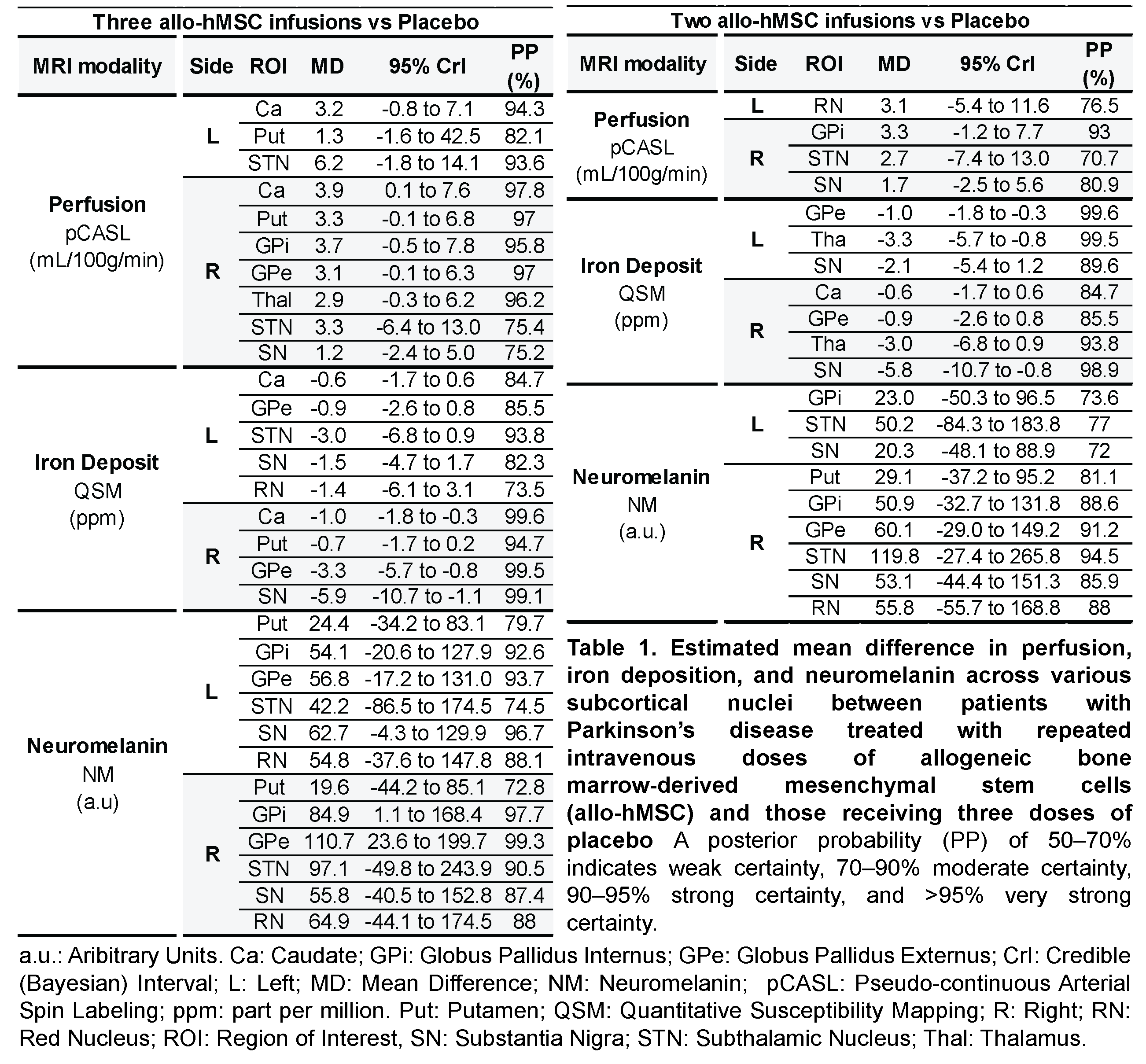
Table 1
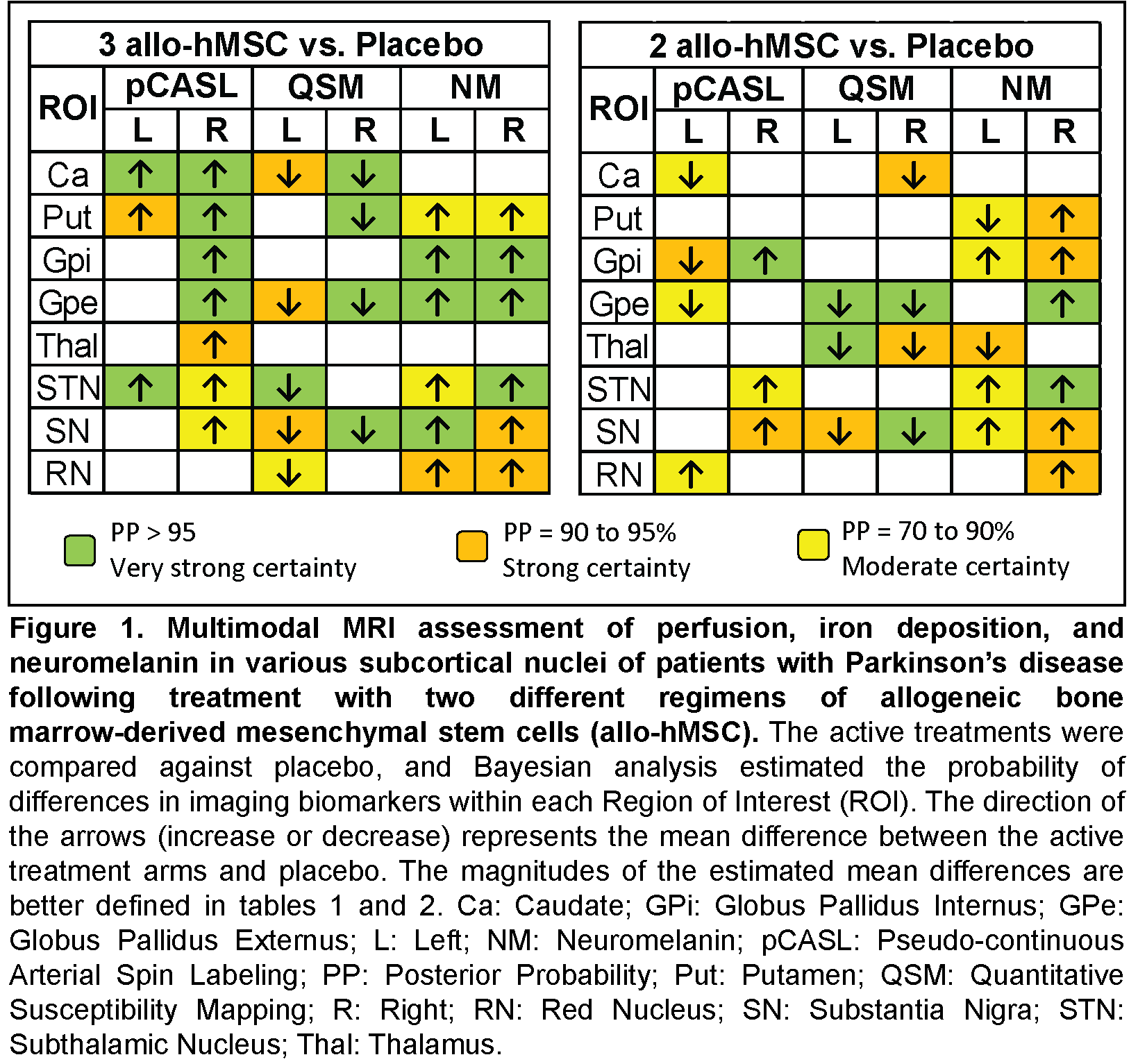
Figure 1
References: [1] Theis H, Pavese N, Rektorová I, van Eimeren T. Imaging Biomarkers in Prodromal and Earliest Phases of Parkinson’s Disease. J Parkinsons Dis. 2024;14(s2):S353-S365. doi: 10.3233/JPD-230385. PMID: 38339941; PMCID: PMC11492013.
[2] Schiess M, Suescun J, Doursout MF, Adams C, Green C, Saltarrelli JG, Savitz S, Ellmore TM. Allogeneic Bone Marrow-Derived Mesenchymal Stem Cell Safety in Idiopathic Parkinson’s Disease. Mov Disord. 2021 Aug;36(8):1825-1834. doi: 10.1002/mds.28582. Epub 2021 Mar 27. PMID: 33772873; PMCID: PMC8451899.
[3] ClinicalTrials.gov. Allogeneic Bone Marrow-Derived Mesenchymal Stem Cell Therapy for Idiopathic Parkinson’s Disea [Internet]. Bethesda (MD): National Library of Medicine (US); 2023 [cited 2025 Feb 26]. Available from: https://clinicaltrials.gov/study/NCT02611167
To cite this abstract in AMA style:
J. Martinez-Lemus, E. Tharp, T. Ellmore, T. Thomas, C. Green, J. Suescun, C. Onuigbo, T. Le, E. Rodarte-Rascon, R. Ritter Iii, M. Schiess. Evaluation of Perfusion, Iron Deposition, and Neuromelanin MRI Biomarkers Following Mesenchymal Stem Cell Treatment in a Phase 2 Parkinson’s Disease Trial [abstract]. Mov Disord. 2025; 40 (suppl 1). https://www.mdsabstracts.org/abstract/evaluation-of-perfusion-iron-deposition-and-neuromelanin-mri-biomarkers-following-mesenchymal-stem-cell-treatment-in-a-phase-2-parkinsons-disease-trial/. Accessed July 10, 2026.« Back to 2025 International Congress
MDS Abstracts - https://www.mdsabstracts.org/abstract/evaluation-of-perfusion-iron-deposition-and-neuromelanin-mri-biomarkers-following-mesenchymal-stem-cell-treatment-in-a-phase-2-parkinsons-disease-trial/