Category: Parkinson's disease: Neuroimaging
Objective: To compare subthalamic nucleus (STN) perfusion changes following mesenchymal stem cell (MSC) infusion in Parkinson’s disease (PD) using Bayesian methods in both Phase I and Phase II trials.
Background: A Phase I trial reported increased STN perfusion six months post-infusion, originally analyzed reporting average STN changes using frequentist methods [1]. A Bayesian re-analysis examined left STN, right STN, and their average, revealing stronger evidence for right STN perfusion increases (BF₁₀=4.245) and bilateral averages (BF₁₀=3.006), but weaker evidence for left STN (BF₁₀ = 0.768). The Phase II study, using a baseline-controlled, between-group analysis, aimed to validate and extend these findings.
Method: Forty-four mild/moderate PD patients were randomized to three groups: three infusions of 10×10⁶ allo-hMSC/kg, one placebo plus two allo-hMSC infusions, or three placebo infusions, at four-month intervals. Participants underwent 3T MRI with pseudo-continuous arterial spin labeling (pCASL) at baseline and one-month post-infusion. Bayesian region-of-interest (ROI) analysis assessed posterior probabilities (PP) and Bayes Factors (BF₁₀) for perfusion changes in left STN, right STN, and their average, controlling for baseline values.
Results: Bayesian analysis in Phase II demonstrated a strong left STN perfusion increase in the 3-infusion group (BF₁₀ = 18.05) but weak evidence for the 2-infusion group (BF₁₀ = 0.273). The right STN showed a strong effect for the 3-infusion group (BF₁₀ = 7.29) and a moderate effect for the 2-infusion group (BF₁₀ = 3.88). These results align with Phase I’s stronger Bayesian evidence for right STN perfusion increases, while the left STN effect became more pronounced in Phase II under repeated dosing and baseline-controlled comparisons.
Conclusion: Phase II replicated and extended the Bayesian evidence from Phase I, showing dose-dependent, lateralized STN perfusion changes. However, methodological differences impact interpretation: Phase I assessed within-subject change (6 months post-infusion vs. baseline), whereas Phase II compared MSC groups to placebo while controlling for baseline values. A stronger right STN effect in Phase I suggests an early perfusion response, while the left STN effect in Phase II may reflect cumulative infusion effects. Bayesian analysis reinforced STN perfusion as a biomarker for MSC therapy in PD.
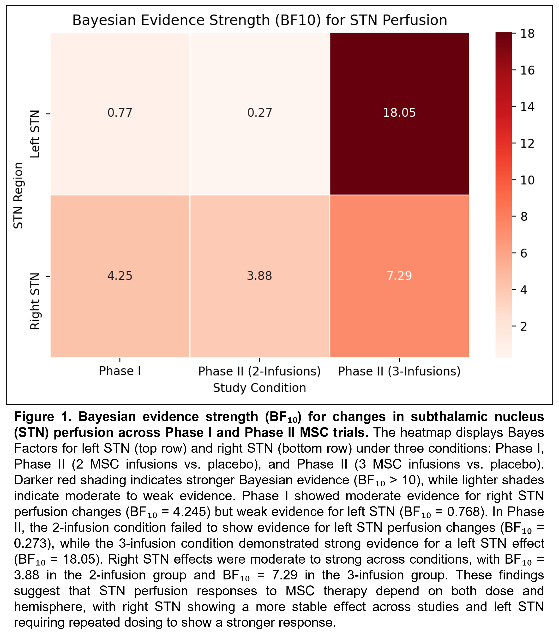
Figure 1
References: [1] Schiess M, Suescun J, Doursout MF, Adams C, Green C, Saltarrelli JG, Savitz S, Ellmore TM. Allogeneic Bone Marrow-Derived Mesenchymal Stem Cell Safety in Idiopathic Parkinson’s Disease. Mov Disord. 2021 Aug;36(8):1825-1834. doi: 10.1002/mds.28582. Epub 2021 Mar 27. PMID: 33772873; PMCID: PMC8451899.
To cite this abstract in AMA style:
T. Thomas, T. Ellmore, E. Tharp, J. Martinez-Lemus, C. Green, J. Suescun, C. Onuigbo, T. Le, E. Rodarte Rascon, R. Ritter Iii, M. Schiess. Bayesian Re-Analysis of Phase I and II Subthalamic Nucleus Perfusion Changes Following Mesenchymal Stem Cell Infusion in Parkinson’s Disease [abstract]. Mov Disord. 2025; 40 (suppl 1). https://www.mdsabstracts.org/abstract/bayesian-re-analysis-of-phase-i-and-ii-subthalamic-nucleus-perfusion-changes-following-mesenchymal-stem-cell-infusion-in-parkinsons-disease/. Accessed July 7, 2026.« Back to 2025 International Congress
MDS Abstracts - https://www.mdsabstracts.org/abstract/bayesian-re-analysis-of-phase-i-and-ii-subthalamic-nucleus-perfusion-changes-following-mesenchymal-stem-cell-infusion-in-parkinsons-disease/