Category: Ataxia
Objective: Describe a young patient with medial past history of Epilepsy who started with progressive symptoms of imbalance, vertigo, nausea, oscillopsy and recurrence of seizures. We suspect an autoimmune movement disorder and perform differential diagnoses included metabolic, vascular, infectious and immuno mediated disorders.
Background: Autoimmune cerebellar ataxia (ACA) is an important and potentially treatable cause of sporadic cerebellar syndrome. These disorders are important in differential diagnoses because they are treatable. Diverse autoimmune-based etiologies, such as gluten ataxia (GA), paraneoplastic cerebellar degeneration (PCD), anti-glutamate decarboxylase 65 antibody-associated cerebellar ataxia (anti-GAD65Ab-associated CA), post-infectious cerebellitis, and opsoclonus myoclonus syndrome (OMS) that account for approximately 6% of all cases. Interestingly, the authors classified 24% of the patients with idiopathic sporadic ataxias.This category included ACA.
Method: A 37-year-old man with history of controlled Epilepsy who suffered from the subacute onset of cerebellar ataxia and uncontrolled seizures. Abnormalities was found in the cerebellum on initial brain MRI (Fig1,2) and we performed lumbar puncture and other neurological test included infectious, vascular and immunological which were negative. Oligoclonal bands were positive and Cerebral Pet showed cerebellar, frontal hypometabolism and focal hypermetabolism suspect thyroid nodules (Fig 3).
Results: His neurological symptoms did improve after intravenous methylprednisolone but significantly improved following Rituximab after 2 months. Electroencephalography showed frontal epileptic activity and endocrinological test was performed prove only benignal nodules and subclinical hypothiroidism. Unfortunately, other antigens could not be performed due to availability in our unit.
Conclusion: Idiopathic sporadic ataxia including ACA are not infrequent however multiple auto-antiboides are involved, however early identification of symptomps and better treatment results in quality of life.
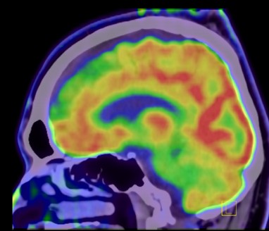
PET bi frontal and cerebellum hypometabolism
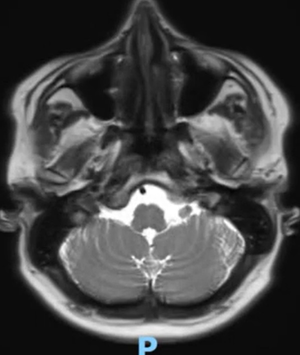
MRI reduction of cerebellum volume in axial T2
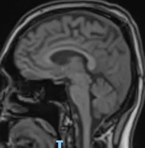
MRI reduction of cerebellum volume in sagital T1
References: 1.Madeline Garza , Amanda L Piquet. Update in Autoimmune Movement Disorders: Newly Described Antigen Targets in Autoimmune and Paraneoplastic Cerebellar Ataxia. Front Neurol. 2021 Aug
2.Balint B. Autoimmune Movement Disorders. Continuum (Minneap Minn). 2024 Aug 1;30(4):1088-1109
3.Hiroshi Mitoma, et al. Immune-mediated Cerebellar Ataxias: Practical Guidelines and Therapeutic Challenges. Curr Neuropharmacol 2019;17(1):33-58
To cite this abstract in AMA style:
J. Balderas Juárez, A. Zepeda Rodríguez, R. Padilla García. A 37 years-old man with Epilepsy and Progressive Autoimmune Ataxia [abstract]. Mov Disord. 2025; 40 (suppl 1). https://www.mdsabstracts.org/abstract/a-37-years-old-man-with-epilepsy-and-progressive-autoimmune-ataxia/. Accessed April 10, 2026.« Back to 2025 International Congress
MDS Abstracts - https://www.mdsabstracts.org/abstract/a-37-years-old-man-with-epilepsy-and-progressive-autoimmune-ataxia/