Category: Parkinson's Disease: Neuroimaging
Objective: To investigate novel cortical diffusivity metrics in Parkinson’s disease (PD), including both prodromal and established genetic PD, alongside sporadic PD and healthy controls (HC).
Background: We have developed metrics of cortical microstructure that relate diffusion tensor imaging (DTI) measures to the minicolumnar direction obtained via modelling the geometry of the cortex: AngleR – the angle of the principal diffusion direction relative to the radial minicolumnar direction; ParlPD – the component of principal diffusion parallel to the minicolumnar direction; PerpPD+ – the components of diffusion perpendicular to the minicolumnar direction. These metrics have shown utility in Alzheimer’s disease [1], frontotemporal lobar degeneration [2] and multiple sclerosis [3]; this work aims to evaluate these imaging biomarkers for PD.
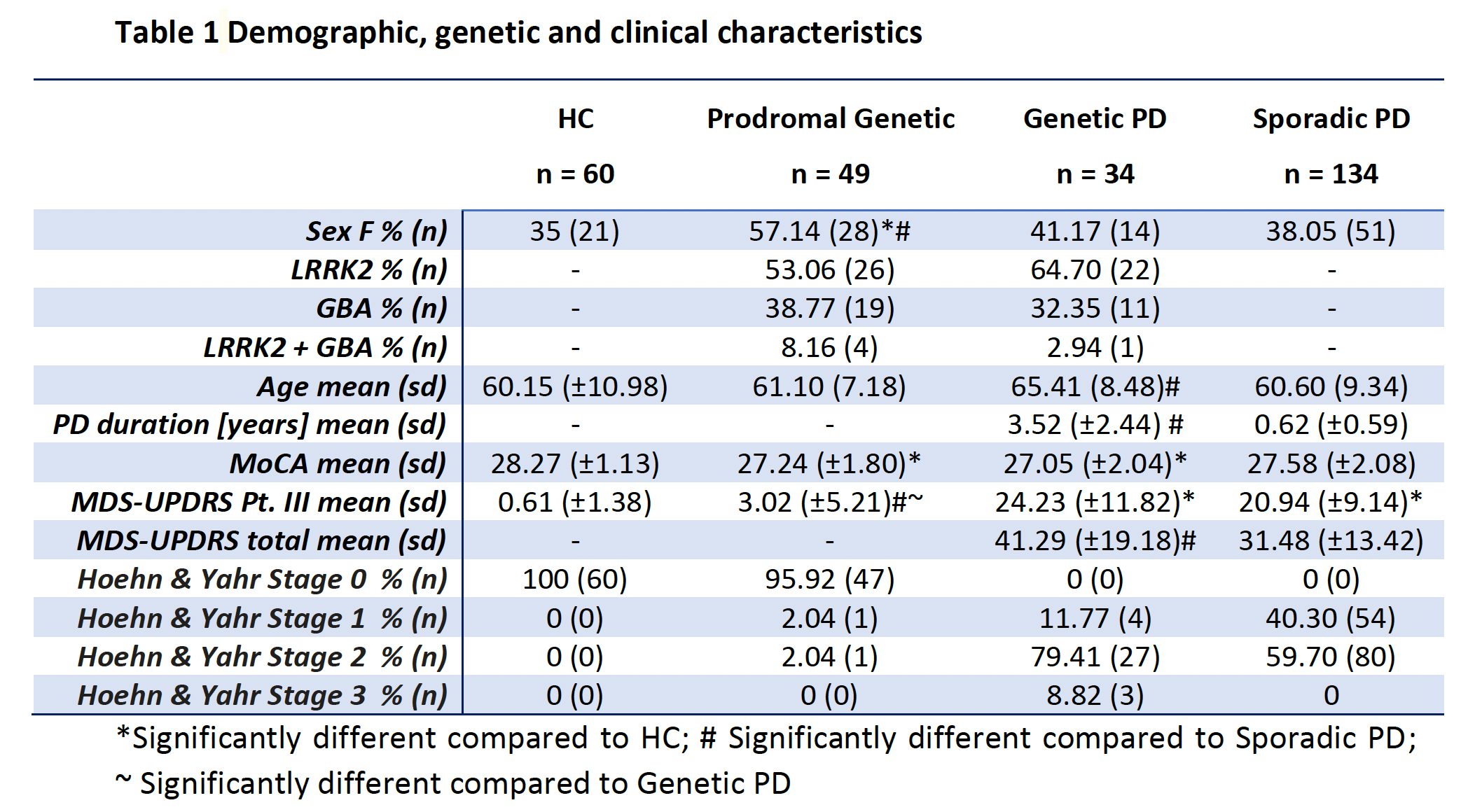
Method: Data was obtained from the Parkinson’s Progression Markers Initiative [4] for 277 participants, detailed in [table1]. 3D T1-weighted structural and DTI scans were analysed to calculate the 3 novel cortical diffusion measures (AngleR, ParlPD and PerpPD+) and the cortical mean diffusivity (MD). Whole brain and regional cortical values were used to investigate cortical microstructural changes along the PD genetic continuum and in sporadic PD. Regional results were considered statistically significant after false discovery rate correction (FDR).
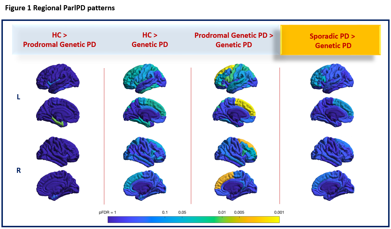
Results: Whole-brain analysis revealed significantly lower ParlPD values in genetic PD, compared to the genetic prodromal group (t264 = 2.75, p = 0.0032). Regional analyses showed a progressive pattern of cortical ParlPD reduction [figure1] across the genetic continuum, with the prodromal results limited to mesocortex, whereas manifest genetic PD also involved neocortex. Comparing the genetic PD and the sporadic PD groups directly, genetic PD showed significantly lower ParlPD in a predominantly frontal pattern. This finding could reflect the significantly higher disease severity reported for the genetic PD group.
Conclusion: Our findings suggest that this novel set of cortical diffusivity metrics is sensitive to the range of cortical architectural changes along the genetic continuum of PD. These results support the use of cortical diffusivity measurements as a marker of cortical microstructural alterations in PD, and warrant further investigation of their correlates and progression over time.
References: [1] Torso, M., Bozzali, M., Zamboni, G., Jenkinson, M., Chance, S. A., & Alzheimers Disease Neuroimage Initiative. (2021). Detection of Alzheimer’s Disease using cortical diffusion tensor imaging. Human brain mapping, 42(4), 967-977.
[2] Torso, M., Ridgway, G. R., Jenkinson, M., & Chance, S. (2021). Intracortical diffusion tensor imaging signature of microstructural changes in frontotemporal lobar degeneration. Alzheimer’s research & therapy, 13(1), 1-15.
[3] McKavanagh, R., Torso, M., … & Chance, S. A. (2019). Relating diffusion tensor imaging measurements to microstructural quantities in the cerebral cortex in multiple sclerosis. Human Brain Mapping, 40(15), 4417-4431.
[4] Data used in the preparation of this article were obtained from the Parkinson’s Progression Markers Initiative (PPMI) database (www.ppmi-info.org/data). For up-to-date information on the study, visit www.ppmi-info.org. PPMI – a public-private partnership – is funded by the Michael J. Fox Foundation for Parkinson’s Research funding partners 4D Pharma, Abbvie, Acurex Therapeutics, Allergan, Amathus Therapeutics, ASAP, Avid Radiopharmaceuticals, Bial Biotech, Biogen, BioLegend, Bristol-Myers Squibb, Calico, Celgene, Dacapo Brain Science, Denali, The Edmond J. Safra Foundaiton, GE Healthcare, Genentech, GlaxoSmithKline, Golub Capital, Handl Therapeutics, Insitro, Janssen Neuroscience, Lilly, Lundbeck, Merck, Meso Scale Discovery, Neurocrine Biosciences, Pfizer, Piramal, Prevail, Roche, Sanofi Genzyme, Servier, Takeda, Teva, UCB, Verily, and Voyager Therapeutics.
To cite this abstract in AMA style:
M. Torso, D. Tzaferou, M. Valotti, J. Hardwidge, I. Hardingham, Q. Guo, R. Comley, S. Chance, G. Ridgway. Assessment of novel diffusion MRI metrics of cortical microstructure in the genetic continuum of Parkinson’s disease [abstract]. Mov Disord. 2022; 37 (suppl 2). https://www.mdsabstracts.org/abstract/assessment-of-novel-diffusion-mri-metrics-of-cortical-microstructure-in-the-genetic-continuum-of-parkinsons-disease/. Accessed May 4, 2026.« Back to 2022 International Congress
MDS Abstracts - https://www.mdsabstracts.org/abstract/assessment-of-novel-diffusion-mri-metrics-of-cortical-microstructure-in-the-genetic-continuum-of-parkinsons-disease/