Session Information
Date: Monday, October 8, 2018
Session Title: Parkinson's Disease: Neuroimaging And Neurophysiology
Session Time: 1:15pm-2:45pm
Location: Hall 3FG
Objective: To identify white matter (WM) changes in PD with neurocognitive-psychiatric symptoms (NPS) using neurite orientation dispersion and density imaging (NODDI) and magnetization transfer saturation (MT-sat) imaging.
Background: Although mainly considered as a movement disorder, NPS are common in PD and have important consequences for quality of life. Sensitive and specific diagnostic biomarkers are necessary for early intervention. NODDI enables more specific characterization of tissue microstructure compared to diffusion tensor imaging by estimating the density and dispersion of neurites. Meanwhile, MT-sat imaging has been shown to be more specific to myelin
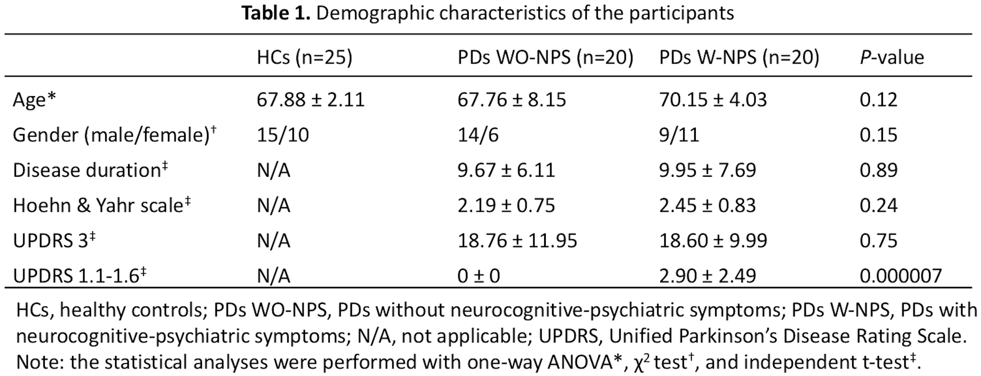
Methods: Forty PD patients (PDs) and 25 healthy controls (HCs) were analyzed (Table 1). PDs were categorized into PD without and with NPS groups where the total of UPDRS 1.1-1.6 equal to 0 (PDs WO-NPS) and ≥ 1 (PDs W-NPS), respectively. Each subject underwent multi-shell diffusion MRI and MT-sat imaging on a 3T MR scanner. Tract-based spatial statistics (TBSS) was used to compare NODDI indices (ICVF, intracellular volume fraction; OD, orientation dispersion index; Viso, isotropic volume fraction) and MT-sat metric (MVF, myelin volume fraction) between HC and PD groups. TBSS was also used to perform correlation analysis on all indices and clinical scores. Further, with ROI analysis the average values of significant indices in the early and late-myelinating tracts were compared between groups and correlated to each other.
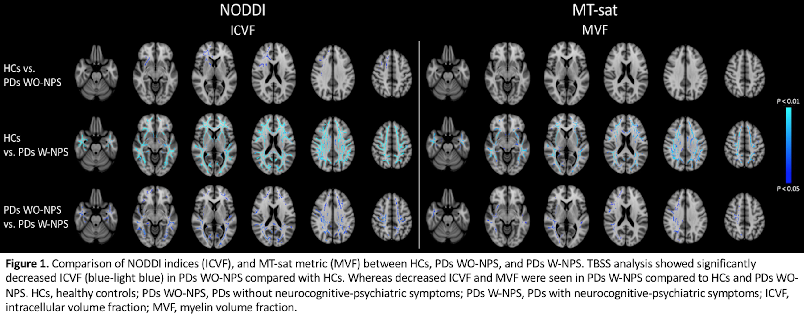
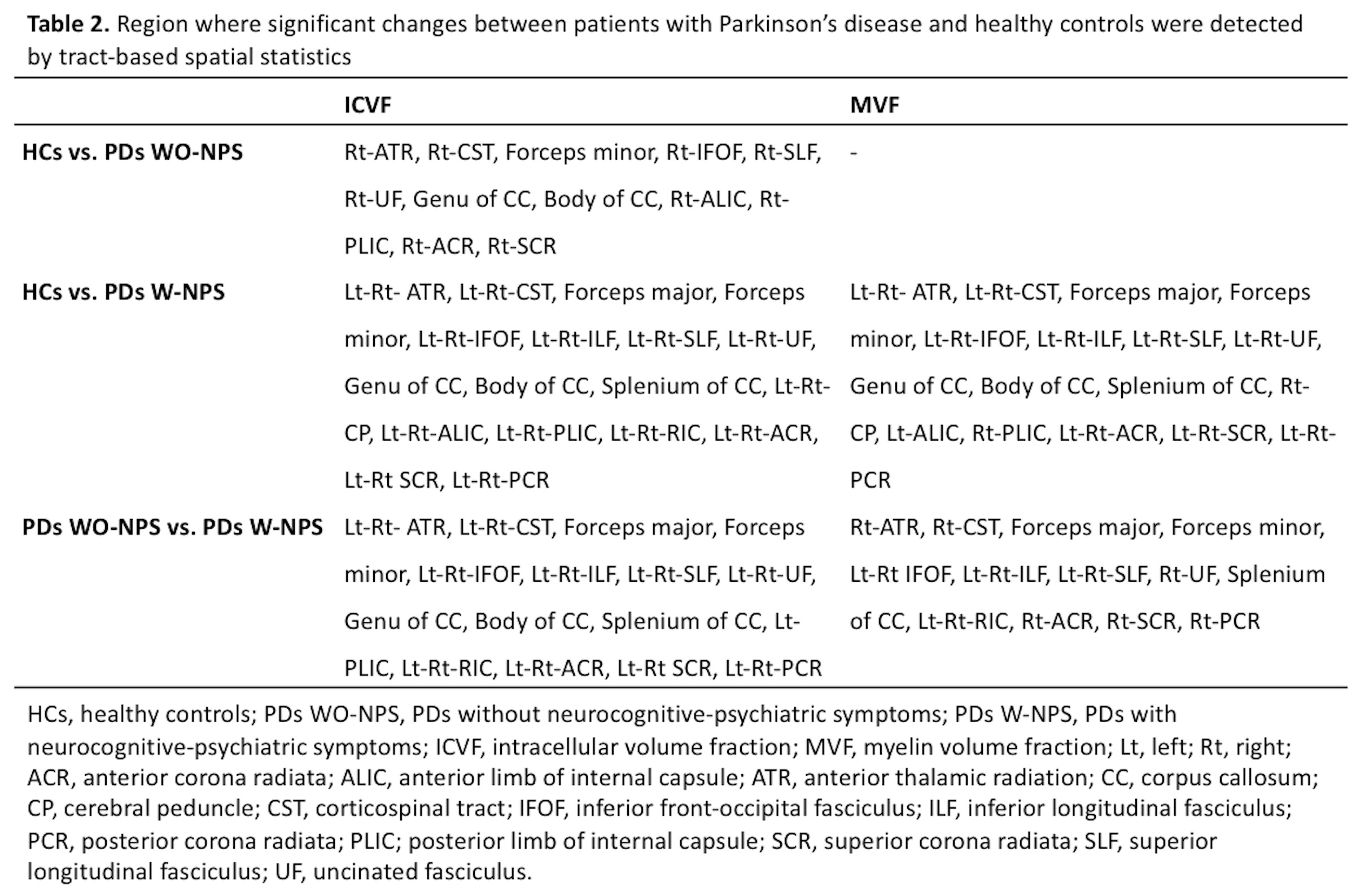
Results: TBSS analysis showed significantly decreased ICVF, which reflects decreased neurite density in the anterior parts of the brain of PDs WO-NPS compared with HCs. In the PDs W-NPS, decreased ICVF and MVF were seen in many WM tracts when compared to HCs or PDs-WO-NPS (Fig. 1, Table 2). No correlations were detected between any of the indices and clinical scores. Further, significant differences of ICVF and MVF values between PDs W-NPS and HCs or PDs WO-NPS were found in the late-myelinating tracts (Fig. 2). Finally, a significant correlation was found between ICVF and MVF in late-myelinating tracts (Fig. 3).
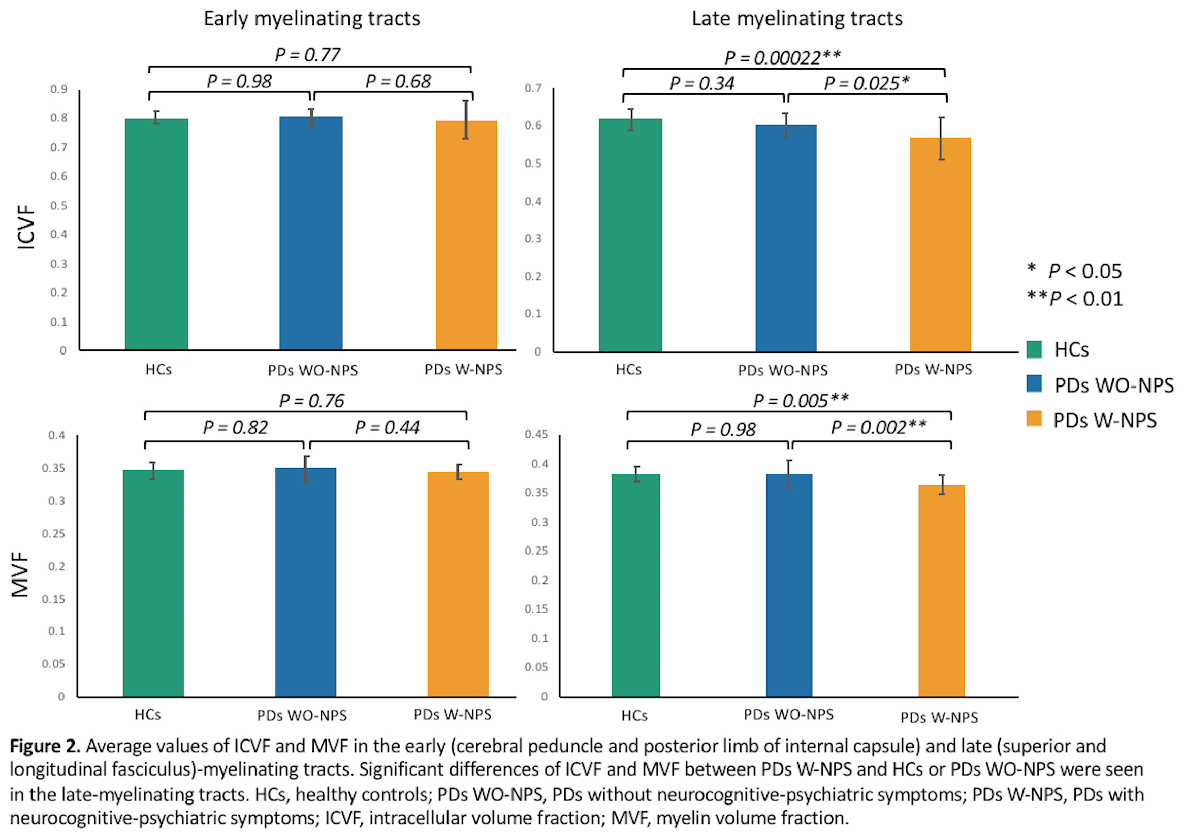

Conclusions: NODDI showed axonal degeneration in the WM of PDs even though the NPS were not yet diagnosed. Our results might also indicate that axonal degeneration precedes and induces demyelination, especially in the late-myelinating tracts. In conclusion, NODDI provides an early biomarker to WM changes in PD with NPS. Whereas combined NODDI and MT-sat imaging can be used to evaluate the disease progression.
To cite this abstract in AMA style:
C. Andica, K. Kamagata, T. Hatano, A. Saito, Y. Takenaka, T. Ogawa, G. Oyama, Y. Shimo, M. Hori, N. Hattori, S. Aoki. Combined Neurite Orientation Dispersion and Density Imaging and Myelin Imaging Provide New Insights to White Matter Changes of the Neurocognitive-psychiatric Disorder in Parkinson’s Disease [abstract]. Mov Disord. 2018; 33 (suppl 2). https://www.mdsabstracts.org/abstract/combined-neurite-orientation-dispersion-and-density-imaging-and-myelin-imaging-provide-new-insights-to-white-matter-changes-of-the-neurocognitive-psychiatric-disorder-in-parkinsons-disease/. Accessed April 9, 2026.« Back to 2018 International Congress
MDS Abstracts - https://www.mdsabstracts.org/abstract/combined-neurite-orientation-dispersion-and-density-imaging-and-myelin-imaging-provide-new-insights-to-white-matter-changes-of-the-neurocognitive-psychiatric-disorder-in-parkinsons-disease/