Category: Ataxia
Objective: To assess changes in brain tissue microstructures using diffusion tensor imaging parameters, fractional anisotropy (FA), increased radial (RD) and axial diffusivities (AD) in SCA 12 patients in comparison with healthy subjects.
Background: Autosomal dominant Spinocerebellar ataxias (SCAs) are one of the most prevalent progressive neurodegenerative ataxias. SCA is characterized by progressive cerebellar ataxia with oculomotor dysfunction, cognitive impairment symptoms pyramidal, and extrapyramidal signs. Diffusion tensor imaging is a quantitative non-invasive method for measuring changes in white matter fibre tracts. Fractional anisotrophy (FA), mean diffusivity (MD), radial diffusivity (RD) and axial diffusivity (AD) are used to study the microstructural damage of white matter.
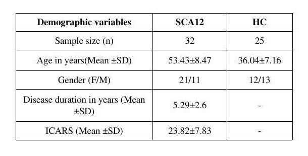
Method: Symptomatic and genetically confirmed SCA12 (M/F:21/11, mean age 53.43±8.47 years) patients and healthy controls (HC, M/F:12/13, mean age 36.04±7.16 years) were recruited after institutional ethical clearance.
MR data was acquired on a 3T MR scanner (Ingenia 3.0 T, M/s. Philips Healthcare, The Netherlands) using a 32-channel head coil. Tl weighted 3D Turbo Field Echo (TFE) sequence (TR/TE: 8.1 /3.7 ms; FOV-240*240; Slices -360 with no gap) was acquired and used as an overlay. Diffusion tensor imaging (DTI) data were acquired using a single-shot echo-planar dual SE sequence in 32 directions with acquisition parameters: b-factor= 0 and 800 s/mm , number of slices=74, slice thickness=2mm, FOV=230 mm×230 mm.
Data processing: Pre and post-processing of the diffusion tensor images was performed using software tools from the FMRIB software library (FSL, http://www.fmrib.ox.ac.uk/fsl). Tract-Based Spatial Statistics (TBSS) were used to create the mean and mean skeletonized map of FA, MD, RD and AD.
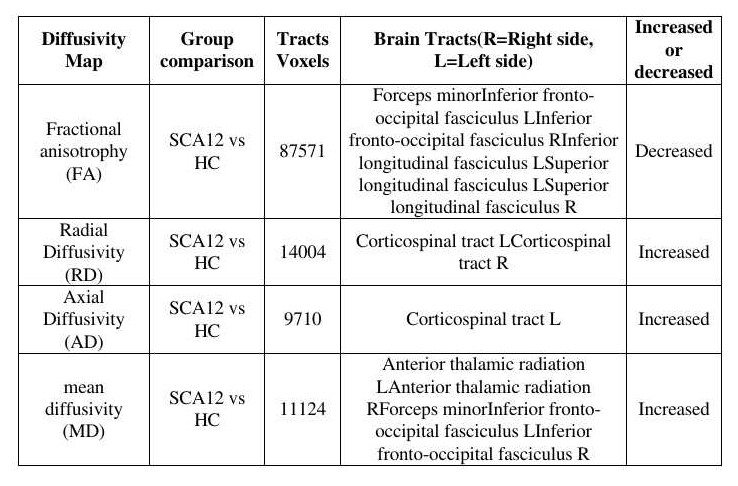
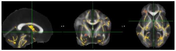
Results: Subject demography is mentioned in table 1. Details of FA values are mentioned in table 2. Bilaterally decreased FA values in the superior and inferior longitudinal fasciculus, anterior thalamic radiation, corticospinal tract were observed in SCA12 groups in comparison with that in healthy controls (Fig 1) .
Conclusion: Atrophy and decreased FA in cerebellum and corticospinal tracts is likely to be associated with motor dysfunction in SCA12. Reduction of FA in major white matter pathways may be associated with cognitive and behavioural impairment.
References: 1. Klockgether, T., Lu, R., Riess, O., Laccone, F., Kramer, B., Abele, M., Bu, K., Scho, L., Boesch, S., Brice, A., Inzelberg, R., Zilber, N., & Dichgans, J. (1998). The natural history of degenerative ataxia : a retrospective study in 466 patients. 589–600.
2. Choudhury, S., Chatterjee, S., Chatterjee, K., Banerjee, R., Humby, J., Mondal, B., Anand, S. S., Shubham, S., & Kumar, H. (2018). Clinical Characterization of Genetically Diagnosed Cases of Spinocerebellar Ataxia Type 12 from India. Movement Disorders Clinical Practice, 5(1), 39–46.
3. Constanzo, J., Dumont, M., Lebel, R., Tremblay, L., Whittingstall, K., Masson-Côté, L., Geha, S., Sarret, P., Lepage, M., Paquette, B., & Descoteaux, M. (2018). Diffusion MRI monitoring of specific structures in the irradiated rat brain. Magnetic Resonance in Medicine, 80(4), 1614–162
To cite this abstract in AMA style:
AK. Srivastava, P. Pankaj, S. Kumaran, A. Garg, R. Agarwal, A. Nehra, F. Mohammad. Diffusion Tensor Imaging of Spinocerebellar ataxia type 12 patients in comparison of Healthy control [abstract]. Mov Disord. 2023; 38 (suppl 1). https://www.mdsabstracts.org/abstract/diffusion-tensor-imaging-of-spinocerebellar-ataxia-type-12-patients-in-comparison-of-healthy-control/. Accessed April 3, 2026.« Back to 2023 International Congress
MDS Abstracts - https://www.mdsabstracts.org/abstract/diffusion-tensor-imaging-of-spinocerebellar-ataxia-type-12-patients-in-comparison-of-healthy-control/