Category: Parkinson's Disease (Other)
Objective: The study aims to compare the connectivity profile of patients affected by sporadic and genetic Parkinson’s disease in different levodopa conditions through the analysis of electroencephalographic (EEG) data.
Background: These two groups of subjects could underlie different functional brain interactions and, consequently, electrophysiological markers. Connectivity analysis allows us to compare the correlation between biological data sources that evolve but are spatially distant, to understand how brain areas talk to each other on a functional rather than anatomical level.
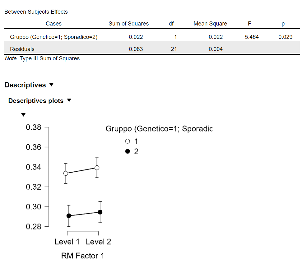
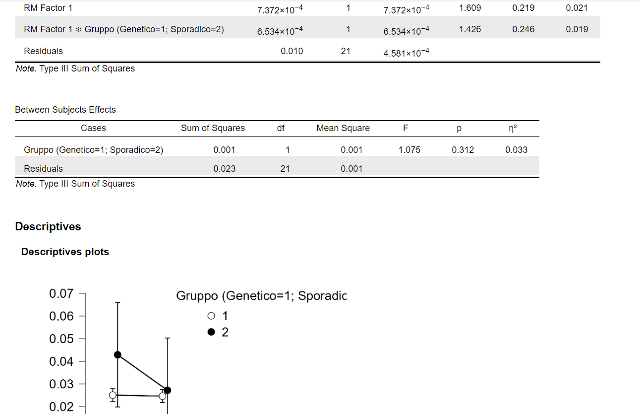
Method: We performed EEG recordings in resting state on a sample of 23 patients with Parkinson’s disease,13 genetic (7 GBA, 6 LRKK2) and 10 sporadic, using the 10-20 standard system in four conditions: ON/OFF therapy, open/closed eyes. After pre-processing through Independent Component Analysis to reduce noise, Functional Connectivity (Imaginary Coherence, Phase-Locking Value) was performed in classical EEG bands of interest (delta, theta, alpha, beta, gamma). A bootstrap approach using phase randomization was applied to assess the statistical significance of non-zero Connectivity values, and to identify valid connectivity nodes. Repeated measures ANOVA was used to compare the ON/OFF connectivity values in the genetic and sporadic patient groups.
Results: Age in the group of genetic patients (52.9 +/- 11) was lower than that of sporadic patients (64 +/- 11, p = 0.03), while Δ UPDRS were comparable (genetic 10.54 +/- 6.15, sporadic 12.89 +/- 8.13, p = 0.45). Connectivity among areas was significantly non-zero in both groups. Medication ON-OFF changes in connectivity in the two groups showed no significant statistical difference. Nor differences in connectivity values were found between patients with genetic and non-genetic Parkinson’s, except for Phase Locking Value analyses in the alpha band. In this analysis, higher connectivity values were found with eyes closed in the genetic patients group compared to the sporadic ones in both MedON and MedOFF, however attributable to the lower average age of the patients in the genetic group.
Conclusion: These results suggest that adaptive and maladaptive phenomena influence brain connectivity in the same way in patients with sporadic or genetic Parkinson’s disease. Additional quantitative linear and non-linear EEG analysis is needed to better characterize these patients’ groups.
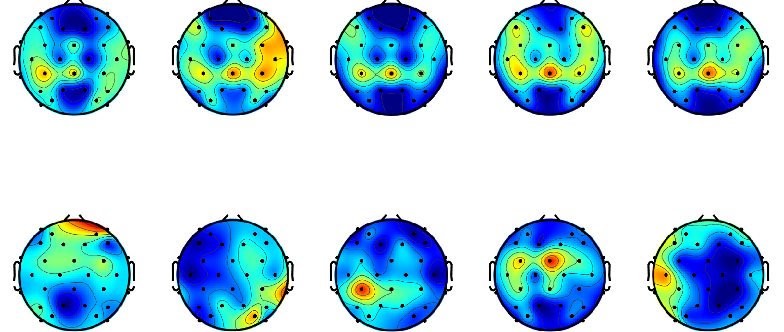
Connectivity in OFF (above PLV; below ImCoh).
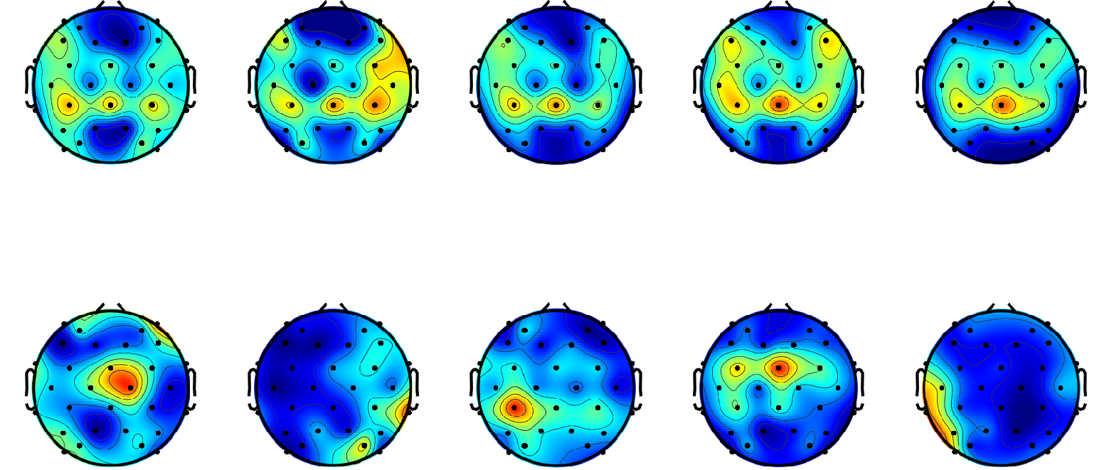
Same patient in ON therapy
PLV alpha band
Imaginary Coherence beta band
References: Karl J Friston – Functional and effective connectivity: a review – Brain
Connect. – 2011 – 1 – 13-36
Fries P. – A mechanism for cognitive dynamics: neuronal communication
through neuronal coherence – Trends Cogn Sci. – 2005 – 9-474
Leviashvili, S.; Ezra, Y.; Droby, A.; Ding, H.; Groppa, S.; Mirelman, A.;
Muthuraman, M.; Maidan, I. – EEG-Based Mapping of Resting-State
Functional Brain Networks in Patients with Parkinson’s Disease
Biomimetics – 2022 – 7 – 231
To cite this abstract in AMA style:
M. Treddenti, V. Yahya, S. Marceglia, V. D'Onofrio, A. Guerra, T. Bocci, A. Priori. EEG connectivity in Parkinson disease: a comparative analysis of sporadic and genetic forms in ON/OFF therapy. [abstract]. Mov Disord. 2025; 40 (suppl 1). https://www.mdsabstracts.org/abstract/eeg-connectivity-in-parkinson-disease-a-comparative-analysis-of-sporadic-and-genetic-forms-in-on-off-therapy/. Accessed April 5, 2026.« Back to 2025 International Congress
MDS Abstracts - https://www.mdsabstracts.org/abstract/eeg-connectivity-in-parkinson-disease-a-comparative-analysis-of-sporadic-and-genetic-forms-in-on-off-therapy/