Category: Parkinson's disease: Neuroimaging
Objective: To evaluate the swallow tail sign in 1.5 Tesla magnetic resonance imaging (MRI) scans of patients diagnosed with Parkinson’s disease (PD).
Background: Nigrosome-1 appears on MRI as a hyperintense structure in the dorsolateral region of the substantia nigra, in a bifurcated shape similar to a swallow’s tail. In PD, the hyperintense signal of nigrosome-1 is lost due to degeneration of dopaminergic neurons, and is described as the loss of the “swallowtail signal”. This concept of nigrosome-1 as a highly sensitive and specific imaging biomarker has been studied extensively in 7 and 3 Tesla MRI, but there are few studies in 1.5 Tesla MRI.
Method: It is a retrospective, single-center study that included PD patients and control patients who underwent brain MRI with susceptibility-weighted imaging (SWI) at 1.5T, independently evaluated by 2 neuroradiologists, as shown in Figure 1.
Results: Thirty-five patients with PD (mean age 66.4±11.5; 29% women) and 12 control patients,who were randomized totaling 36 samples (mean age 63.4±10.4; 67% women), were included (Table 1). The interobserver agreement for the evaluation of the swallow tail sign between the radiologists was moderate (kappa = 0.55). The consensus between the evaluators, in regards to the loss of the swallow tail sign, showed a sensitivity of 70%, specificity of 83%, positive predictive value of 81%, negative predictive value of 73% and accuracy of 67% (Table 2).
Conclusion: The swallow tail sign has been shown to be useful in the diagnosis of PD and, despite superior results in 3T fields, it is possible to consider evaluation in 1.5 T MRI. Our study showed promising results, but has some limitations. Multicenter studies with a larger number of participants, with different MRI equipment (1.5T and 3T) and with training of neuroradiologists should be performed in the future in order to define the diagnostic accuracy of the swallow tail sign in MRI of smaller fields.
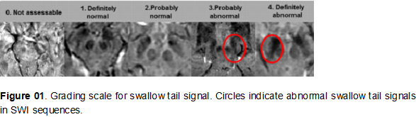
Figure 1
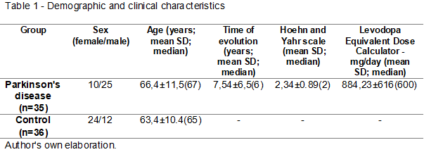
Table 1
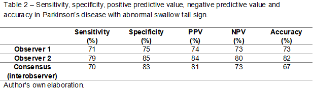
Table 2
To cite this abstract in AMA style:
E. Vioncek, M. Ferreira Cordellini, L. Kami, B. Teixeira. Evaluation of swallow tail sign on 1.5 Tesla Magnetic Resonance Imaging in Parkinson´s disease patients in a tertiary hospital [abstract]. Mov Disord. 2025; 40 (suppl 1). https://www.mdsabstracts.org/abstract/evaluation-of-swallow-tail-sign-on-1-5-tesla-magnetic-resonance-imaging-in-parkinsons-disease-patients-in-a-tertiary-hospital/. Accessed April 7, 2026.« Back to 2025 International Congress
MDS Abstracts - https://www.mdsabstracts.org/abstract/evaluation-of-swallow-tail-sign-on-1-5-tesla-magnetic-resonance-imaging-in-parkinsons-disease-patients-in-a-tertiary-hospital/