Category: Parkinson's Disease: Disease mechanisms
Objective: To investigate plasma neuron-derived ferritin-containing extracellular vesicles (NDFEVs) differences between Parkinson’s disease (PD) patients and controls, assess their association with brain iron deposition, and explore the role of α-synuclein (α-syn) in ferritin-related brain iron deposition in PD.
Background: Iron dysregulation contributes to the pathogenesis of PD. Ferritin, a crucial iron-binding protein, has been reported significantly reduced in the substantia nigra (SN) of PD patients. α-Syn, the main component of Lewy bodies, may be involved in iron metabolism. However, the mechanisms by which α-syn influences ferritin-related iron accumulation in the brain, and its involvement in the PD pathology, remain unclear.
Method: We developed a nanoscale flow cytometry assay to assess the NDFEVs in plasma from patients with PD, multiple system atrophy (MSA), isolated REM behavior disorder (iRBD) and healthy controls (HCs). Ferritin levels in neuron-derived EVs (NDEVs) were assessed by the EV immunoprecipitation and a chemiluminescent immunoassay. Iron deposition in brain was evaluated by quantitative susceptibility mapping (QSM). Human brain slices and SH-SY5Y cells were employed to study the role of α-syn in the regulation of ferritin and iron deposition.
Results: The level of NDFEVs was significantly lower in PD patients compared to HCs (AUC = 0.83) and MSA patients (AUC = 0.80). Ferritin levels in NDEVs were also significantly reduced in PD patients compared to HCs. An integrated model combining ferritin levels in NDEVs and NDFEVs improved the AUC to 0.88. In PD patients, plasma NDFEVs concentration exhibited negative correlations with the MDS-UPDRS III (R = – 0.300; p = 0.004). QSM analysis revealed significantly higher QSM values in the substantia nigra pars compacta (SNpc) of PD patients compared to HCs. The QSM value of the SNpc in PD patients negatively correlated with NDFEVs (R = – 0.263, p = 0.042). Additionally, α-syn significantly reduced ferritin levels in SH-SY5Y cells and decreased the release of ferritin-containing EVs. Finally, our findings suggest that α-syn promotes ferritin degradation through the ubiquitin-proteasome pathway, with Itch mediating this process.
Conclusion: Peripheral NDFEVs may serve as a diagnostic biomarker for PD, reflecting iron deposition in the SNpc. α-Syn pathology is involved in Itch-mediated ferritin degradation and brain iron accumulation in PD.
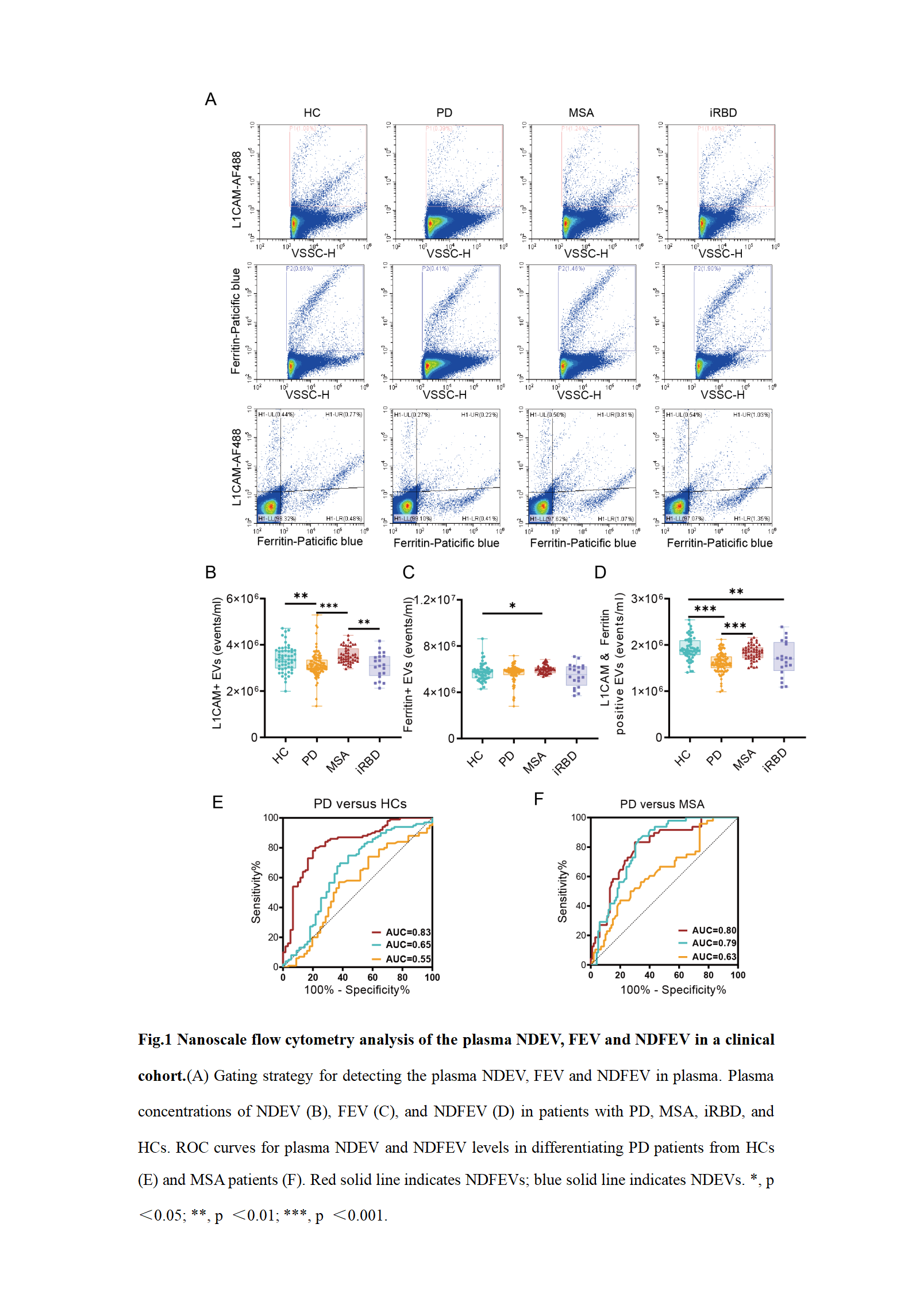
fig.1
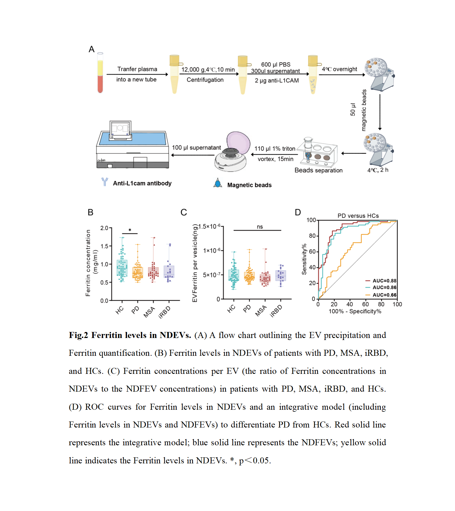
fig.2
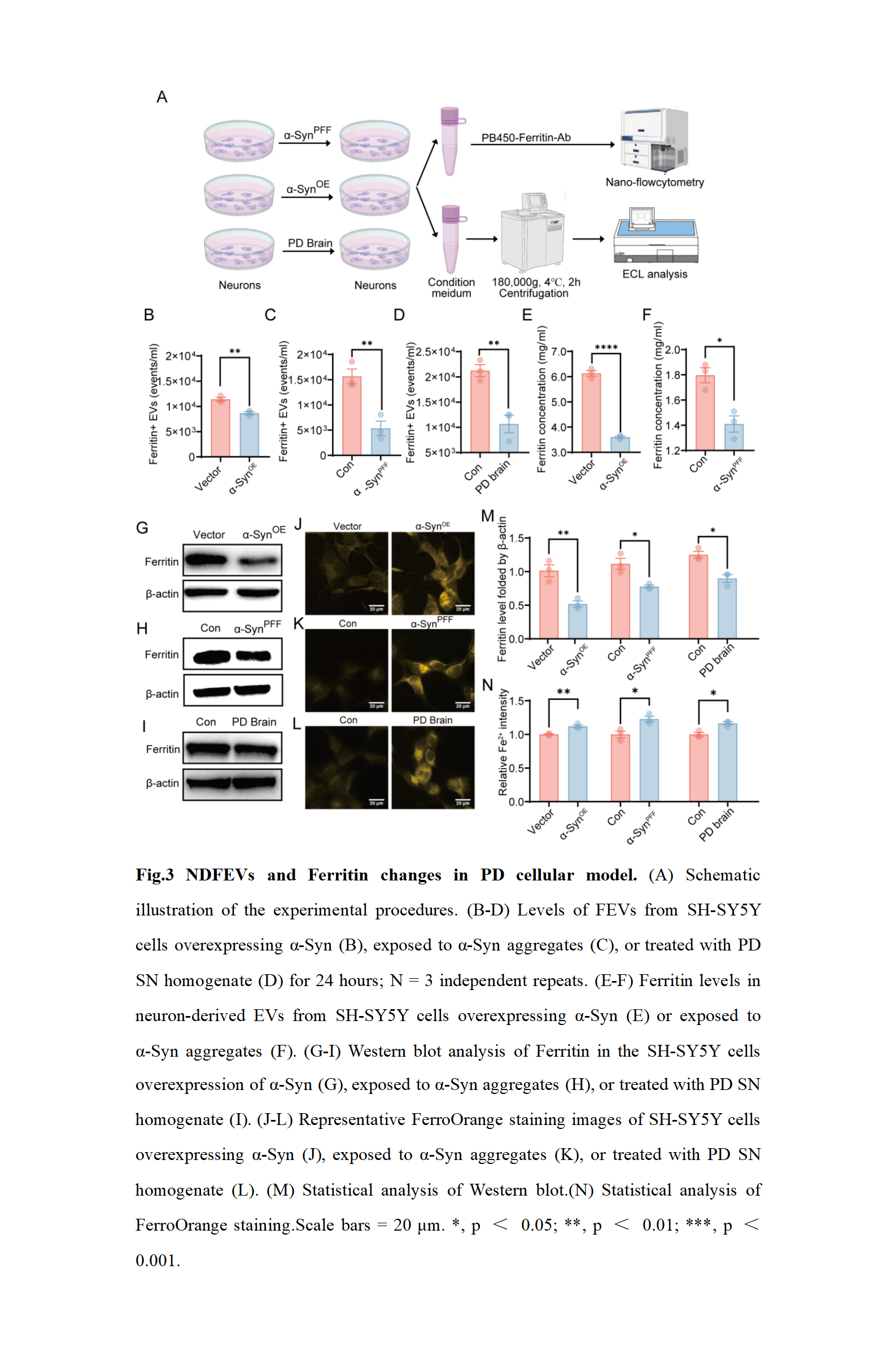
fig.3
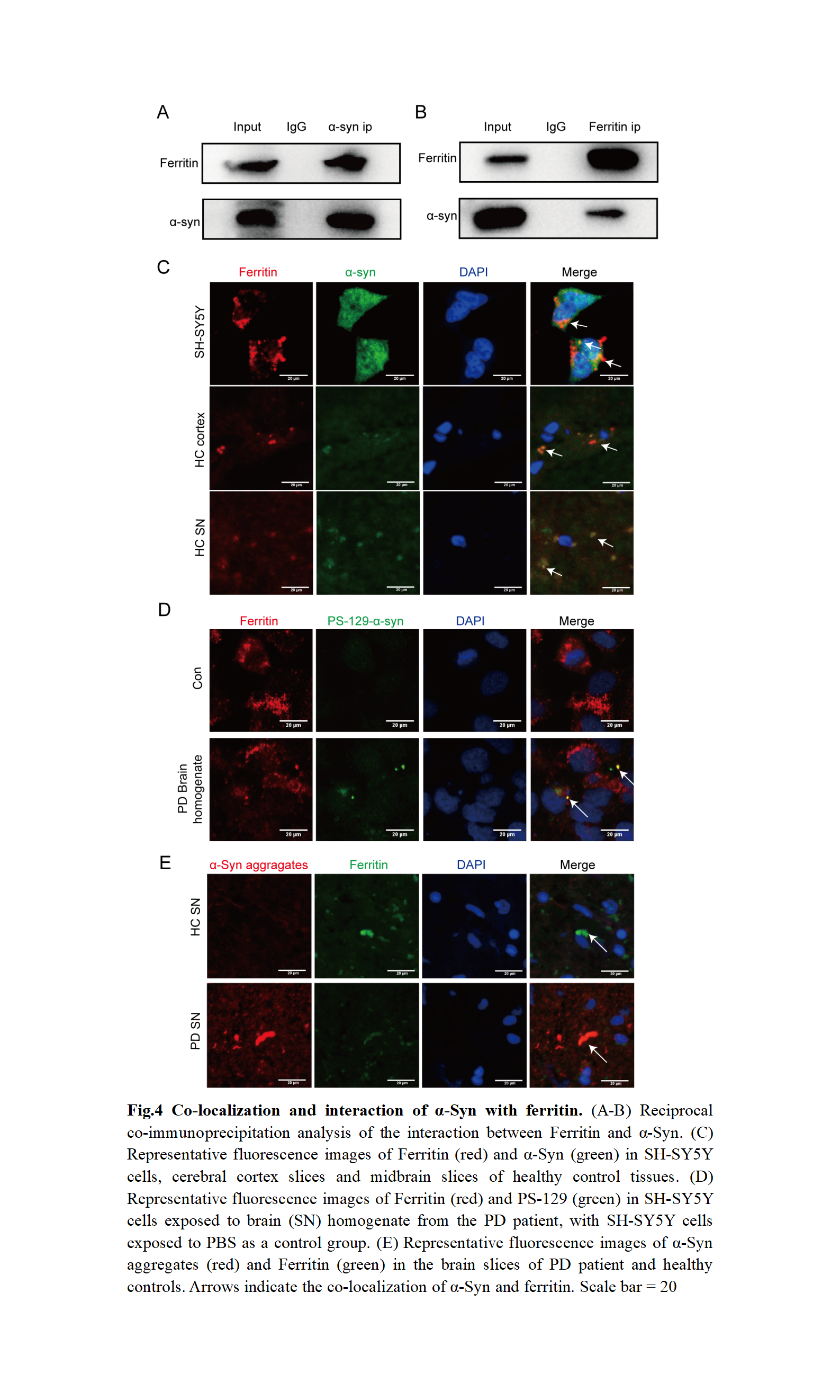
fig.4
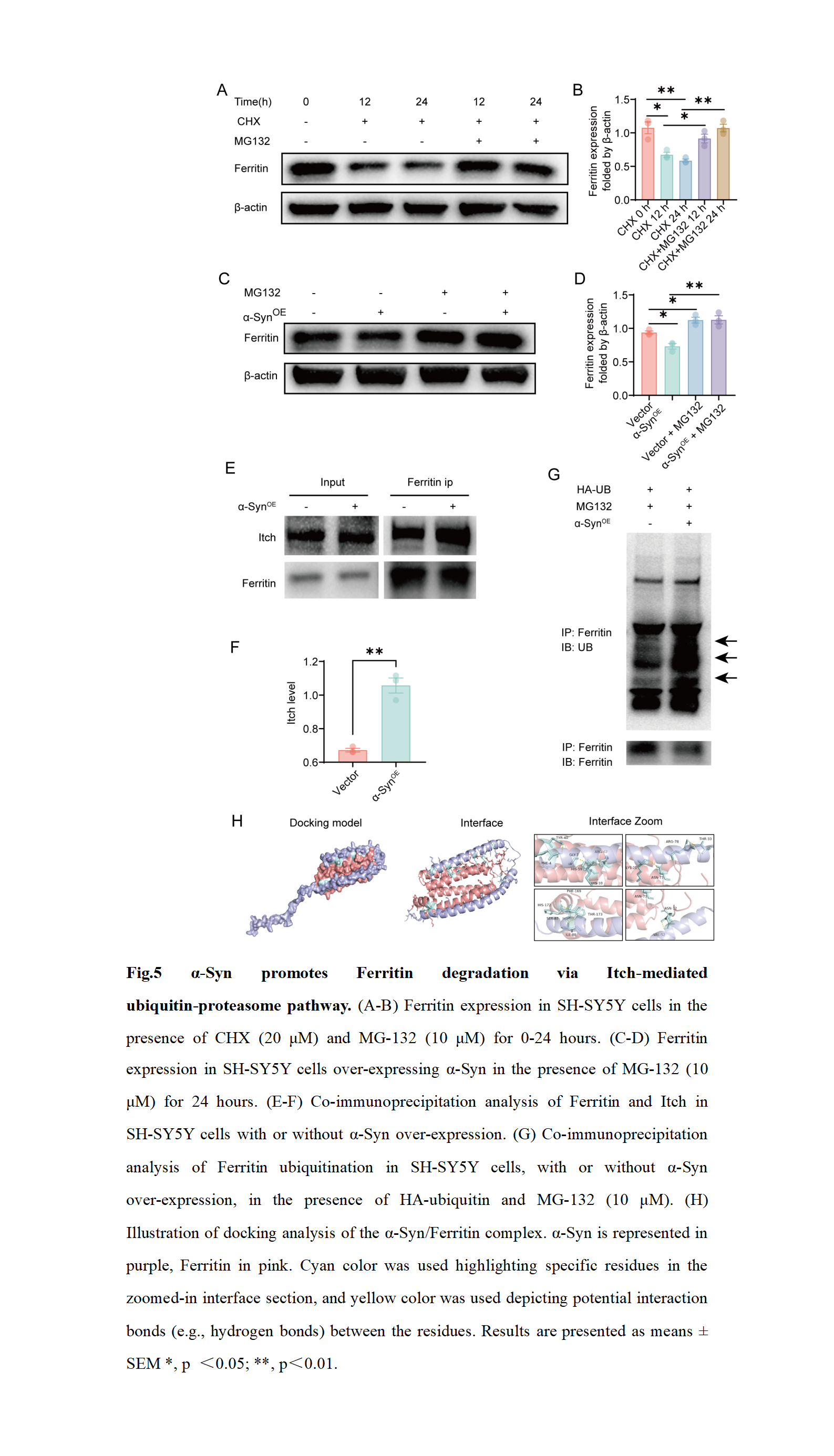
fig.5
To cite this abstract in AMA style:
L. Luo, D. Su, Z. Yu, T. Feng. Ferritin-related Brain Iron Deposition in Parkinson’s Disease Involves α-Synuclein Pathology [abstract]. Mov Disord. 2025; 40 (suppl 1). https://www.mdsabstracts.org/abstract/ferritin-related-brain-iron-deposition-in-parkinsons-disease-involves-%ce%b1-synuclein-pathology/. Accessed April 11, 2026.« Back to 2025 International Congress
MDS Abstracts - https://www.mdsabstracts.org/abstract/ferritin-related-brain-iron-deposition-in-parkinsons-disease-involves-%ce%b1-synuclein-pathology/