Objective: This study aims to classify healthy control (HC) participants, pre-symptomatic Spinocerebellar Ataxia Type 3 (SCA3) patients (preSCA3), and symptomatic SCA3 patients (SCA3) by processing the brainstem surface using a graph neural network (GNN). Subsequently, an explainable AI (XAI) approach is used to identify brainstem regions crucial for classification, which are further statistically analyzed to uncover potential biomarkers of SCA3 pathology.
Background: Spinocerebellar Ataxia Type 3 (SCA3) involves brainstem and cerebellar atrophy. So far, no treatment is available, but the first gene therapy trials have started recently. Thus, there is an urgent need for non-invasive, quantitative biomarkers.
Method: Participants from the ESMI cohort underwent MRI assessments, and T1-weighted images were obtained alongside clinical symptom scores. Brainstem surfaces were reconstructed, triangulated into meshes, and transformed into graphs to train a GNN. The reported scores were calculated by leaving out 20% of subjects for testing. An XAI model identified key regions for classification, and features derived from these regions were statistically analyzed. Significant biomarkers were identified after correction for multiple comparisons and accounting for age, sex, and estimated total intracranial volume (eTIV). The methodology is depicted in Fig. 1.
Results: In 167 participants (368 T1-weighted scans: 111 HC, 73 preSCA3, 184 SCA3), GNN classification achieved high ROC-AUC scores: HC vs. preSCA3 (97%, PR-AUC: 94%), HC vs. SCA3 (97%, PR-AUC: 99%), and preSCA3 vs. SCA3 (94%, PR-AUC: 98%). The XAI model assigned high importance to the medulla oblongata. Statistical analyses revealed significant group differences in multiple features, including the surface area (p<0.01 for both HC vs. preSCA3 and HC vs. SCA3) and the largest 3D diameter (p<0.01 for both HC vs. preSCA3 and HC vs. SCA3).
Conclusion: This study demonstrated that GNNs can effectively model brainstem shape alterations in SCA3. The XAI approach successfully identified key regions showing significant differences between groups both in the early stages (HC vs. preSCA3) and in the later, symptomatic stages of the disease (HC vs. SCA3). The presented approach of AI-guided biomarker discovery, generalizable to other conditions, can accelerate the identification of biomarkers in SCA3 and other neurological disorders.
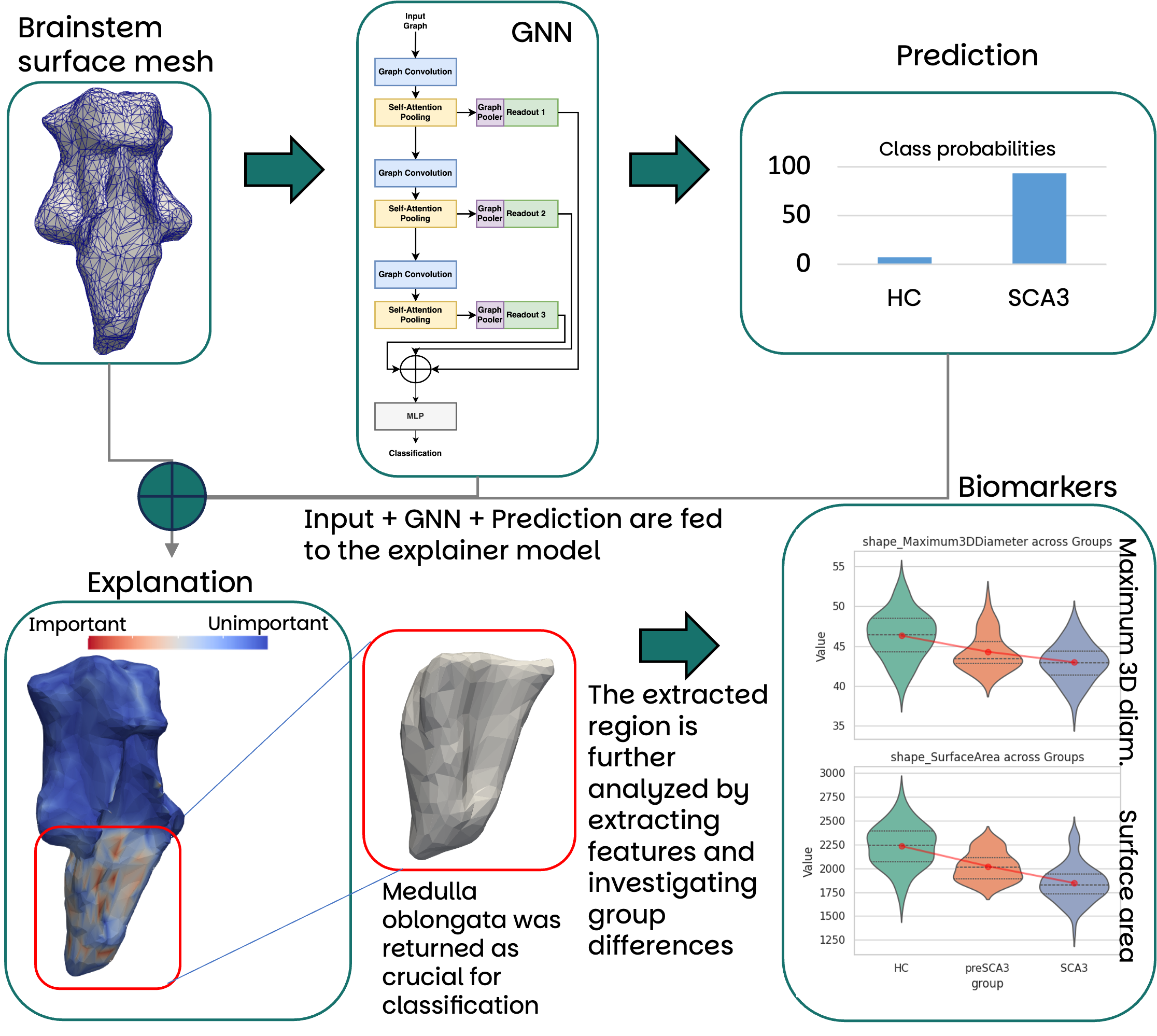
Figure 1
To cite this abstract in AMA style:
P. Wegner, M. Ferreira, J. Theisen, T. Klockgether, J. Faber. From Explainable AI to Biomarkers: Identifying Disease-Related Brain Regions in Spinocerebellar Ataxia Type 3 [abstract]. Mov Disord. 2025; 40 (suppl 1). https://www.mdsabstracts.org/abstract/from-explainable-ai-to-biomarkers-identifying-disease-related-brain-regions-in-spinocerebellar-ataxia-type-3/. Accessed April 7, 2026.« Back to 2025 International Congress
MDS Abstracts - https://www.mdsabstracts.org/abstract/from-explainable-ai-to-biomarkers-identifying-disease-related-brain-regions-in-spinocerebellar-ataxia-type-3/