Category: MSA, PSP, CBS: Neuroimaging
Objective: This study investigated functional brain network alterations in early-stage Parkinson’s disease (PD) and multiple system atrophy (MSA) patients with probable rapid eye movement sleep behavior disorder (pRBD), aiming to differentiate disease-specific changes and assess their biomarker potential.
Background: RBD is a common non-motor symptom observed in synucleinopathies. However, functional brain network alterations associated with RBD in these disorders and differences between PD and MSA patients with pRBD remain insufficiently explored.
Method: A total of 207 individuals were included: 23 PD-pRBD+, 52 PD-pRBD-, 32 MSA-pRBD+, 49 MSA-pRBD-, and 51 healthy controls (HC). Functional connectivity (FC) was assessed via independent component analysis of resting-state fMRI. Receiver operating characteristic (ROC) analysis and area under the curve (AUC) were used to evaluate the diagnostic potential of FC alterations.
Results: PD and MSA patients with pRBD exhibited greater disease severity. PD-pRBD+ patients exhibited reduced FC within the dorsal attention network (DAN) compared to both HC and PD-pRBD- patients, and increased FC between the basal ganglia (BG) and both the sensorimotor network and salience network (SN), as well as between the auditory network (AN) and right frontoparietal network (FPN) compared to HC (Figure 1A). MSA-pRBD+ patients showed widespread intra-network FC reductions, and increased FC between the anterior default mode network (aDMN) and left FPN compared to HC, along with altered FC within the medial visual networks (mVN) and between the mVN and left FPN compared to MSA-pRBD- patients (Figure 1B). Direct comparisons between PD-pRBD+ and MSA-pRBD+ patients revealed decreased FC within the DAN in PD-pRBD+, whereas MSA-pRBD+ patients demonstrated reduced FC within the mVN and inter-network FC in SN-BG, and AN-aDMN (Figure 1C). ROC analysis showed that combined FC values reached high AUC for differentiating PD-pRBD+ and MSA-pRBD+ from HC. Notably, the combination of FC within the DAN, mVN, and FC between the SN and BG, achieved an AUC of 0.94 for distinguishing MSA-pRBD+ from PD-pRBD+.
Conclusion: PD and MSA patients with pRBD exhibited distinct FC alterations, providing insights into disease-specific mechanisms. Combined FC alterations demonstrated high discriminative power and may serve as valuable biomarkers for the early differential diagnosis of these conditions.
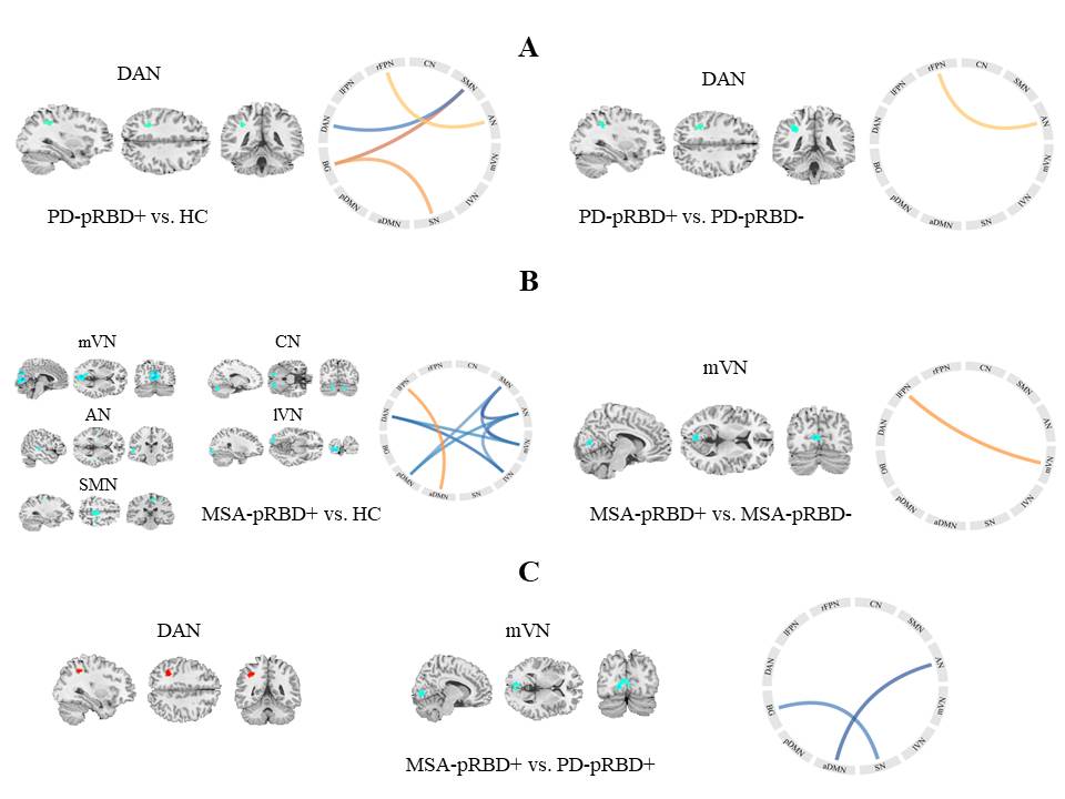
Figure 1
To cite this abstract in AMA style:
SC. Wang, Y. Xiao, YB. Hou, CY. Li, RW. Ou, JY. Lin, TM. Yang, NN. Che, QR. Jiang, XT. Zheng, JY. Liu, HF. Shang. Functional Alterations in Early-Stage Parkinson’s Disease and Multiple System Atrophy with REM Sleep Behavior Disorder [abstract]. Mov Disord. 2025; 40 (suppl 1). https://www.mdsabstracts.org/abstract/functional-alterations-in-early-stage-parkinsons-disease-and-multiple-system-atrophy-with-rem-sleep-behavior-disorder/. Accessed April 10, 2026.« Back to 2025 International Congress
MDS Abstracts - https://www.mdsabstracts.org/abstract/functional-alterations-in-early-stage-parkinsons-disease-and-multiple-system-atrophy-with-rem-sleep-behavior-disorder/