Session Information
Date: Wednesday, September 25, 2019
Session Title: Neuroimaging
Session Time: 1:15pm-2:45pm
Location: Les Muses Terrace, Level 3
Objective: To unravel functional connectivity (FC) traits in Parkinson’s disease mild cognitive impairment (PD-MCI).
Background: PD-MCI is a well-known risk factor for PD associated dementia. Several FC changes have been previously reported (1). Two connectome analyses were used: Network Based analysis (NBS) and Connectome Independent Component Analysis (connICA).
Method: 23 PD-MCI patients, 21 cognitively normal PD (PDCN) patients, and 21 healthy controls (HC) were scanned in a 3T MR scanner. PD-MCI was diagnosed according to the MDS Task Force Guidelines (level II). T1-w and T2-w high resolution scans, and 10 minutes eyes-open resting state BOLD fMRI images with standard (monoband, TR=2s) and multiband (TR=800ms) images were obtained. MRI preprocessing included: T1-w parcellation into 72 cortical and 8 subcortical regions for hemisphere (Freesurfer), standard fMRI pre-processing, and nuisance regression (AFNI). Next, we computed FC matrices for multiband and monoband images for NBS (2) and connICA (3). F values were computed with mixed random effect model for each FC-trait weight to reveal significant differences between groups.
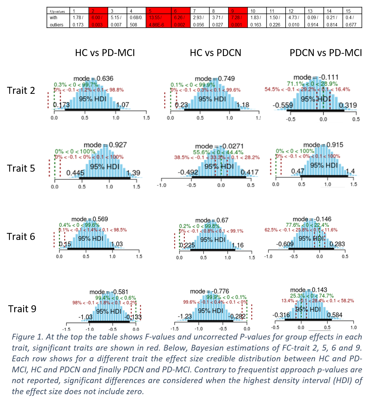
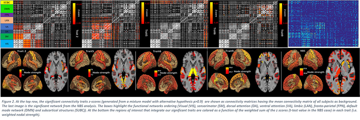
Results: Four FC-traits showed significant differences across groups (Table in [figure1]) (p ≤ 0.05/15). FC-traits 2, 6 and 9 showed significant differences in the comparisons HC vs. PDCN and HC vs. PD-MCI. In FC-trait 5 a significant effect size was found for HC vs. PD-MCI and PDCN vs PD-MCI [figure1]. FC-trait 2 encompasses the visual (VN), the sensorimotor (SMN), and the attention networks (AN). FC-trait 6 represents the connectivity of cortical structures with the thalamus. And FC-trait 9 mirrors connections of the Default Mode Network (DMN) with other cortical regions [figure2]. A unique trait (FC-trait 5) differentiates PD-MCI patients from the other groups. FC-trait 5 includes connections between the SMN, limbic networks, and, mainly the AN, whose disruptions have been previously described in PD-MCI patients. The NBS analysis showed differences among PDCN, PD-MCI, and HC in a network that resembles the combination of FC-traits 2 and 5.
Conclusion: Three independent FC-traits encompassing networks involving cognitive and motor functions were found in PD versus HC. Interestingly, a unique FC-trait encompassing the SMN, attentional networks and limbic networks was the fingerprint of PD-MCI patients. This FC-trait might be explored in future studies as a biomarker for PD-MCI.
References: 1. Wolters, A. F. et al. Resting-state fMRI in Parkinson’s disease patients with cognitive impairment: A meta-analysis. Parkinsonism & Related Disorders 0, (2018). 2. Zalesky, A., Fornito, A. & Bullmore, E. T. Network-based statistic: identifying differences in brain networks. Neuroimage 53, 1197–1207 (2010). 3. Amico, E. et al. Mapping the functional connectome traits of levels of consciousness. Neuroimage 148, 201–211 (2017).
To cite this abstract in AMA style:
M. Delgado-Alvarado, V. Ferrer-Gallardo, I. Navalpotro-Gómez, S. Moia, M. Carreiras, C. Caballero-Gaudes, MC. Rodríguez-Oroz. Functional connectome in Parkinson’s disease mild cognitive impairment [abstract]. Mov Disord. 2019; 34 (suppl 2). https://www.mdsabstracts.org/abstract/functional-connectome-in-parkinsons-disease-mild-cognitive-impairment/. Accessed March 22, 2026.« Back to 2019 International Congress
MDS Abstracts - https://www.mdsabstracts.org/abstract/functional-connectome-in-parkinsons-disease-mild-cognitive-impairment/