Category: Parkinson's Disease: Surgical Therapy
Objective: To develop a therapeutic system to allow real time modulation of striatal dopamine release using a living electrode composed of optogenetically controlled dopaminergic neurons. The first step of which is to optimize the transduction efficiency of human induced pluripotent stem cell (iPSC)-derived dopaminergic neurons for optical control and cell health.
Background: Unfortunately, gold standard PD treatments, like dopamine replacement therapy and deep brain stimulation (DBS), can lose efficacy, are nonspecific, and do not replace dopaminergic neurons. We are developing a strategy to control striatal dopamine levels featuring implantable, living axonal tracts projecting from a discrete population of optically controlled dopaminergic neurons called Dopaminergic Tissue Engineered Living Electrodes (dTELEs) [Figure1][1,2].
Method: Following aggregation, iPSC-derived dopaminergic neurons underwent viral transduction with AAV1 and AAV2 vectors carrying fluorophores and Channelrhodopsin 2. They were seeded into hydrogel microcolumns to fabricate dTELEs or cultured on a 2D surface. Phase-contrast images were taken over time to assess neuronal health and axonal outgrowth. Cultures were imaged live using confocal microscopy to determine vector transduction and phenotype. Standard viability assays were used to ascertain health.
Results: iPSC-derived dopaminergic neurons can be successfully transduced using AAV channelrhodopsins. Groups treated with a fluorophore-only vector or higher vector titers had improved transduction efficiency. Some groups’ cell viability and health metrics declined with increased viral titers, while health metrics differed between vectors at matching titers. Ongoing studies are evaluating the effects of viral transduction on cellular health and viability, mitochondrial health, integration with medium spiny neurons, and evoked dopamine release with optical stimulation.
Conclusion: We are developing a tissue engineering-based treatment for PD that replaces lost dopaminergic neurons, directs axonal growth and connection to the striatum, and allows for specific, controlled external activation to regulate striatal dopamine levels. We anticipate that dTELEs will serve as the foundation of a paradigm-shifting PD therapy that enables tailor-made treatments to address the root cause of PD motor symptoms rather than the symptoms themselves.
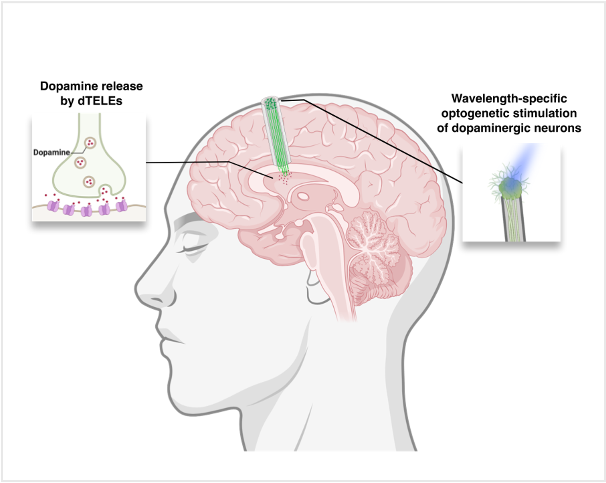
Dopaminergic Tissue Engineered Living Electrodes
References: [1] Dayo O. Adewole et al., Development of Optically Controlled “Living Electrodes” With Long-Projecting Axon Tracts for A Synaptic Brain-Machine Interface. Sci. Adv. 7, eaay5347 (2021). DOI: 10.1126/sci adv.aay5347
[2] Struzyna, Laura A et al. “Tissue Engineered Nigrostriatal Pathway for Treatment of Parkinson’s Disease.” Journal Of Tissue Engineering and Regenerative Medicine vol. 12,7 (2018): 1702-1716. doi:10.1002/term.2698
To cite this abstract in AMA style:
K. Yankson, D. Chouhan, J. Duda, K. Cullen. Living Deep Brain Stimulation for External Control of Striatal Dopamine [abstract]. Mov Disord. 2025; 40 (suppl 1). https://www.mdsabstracts.org/abstract/living-deep-brain-stimulation-for-external-control-of-striatal-dopamine/. Accessed April 10, 2026.« Back to 2025 International Congress
MDS Abstracts - https://www.mdsabstracts.org/abstract/living-deep-brain-stimulation-for-external-control-of-striatal-dopamine/