Session Information
Date: Sunday, October 7, 2018
Session Title: Parkinsonism, MSA, PSP (Secondary and Parkinsonism-Plus)
Session Time: 1:45pm-3:15pm
Location: Hall 3FG
Objective: To establish whether high-resolution computer tomography (CT) scans can aid the diagnosis of progressive supranuclear palsy (PSP), and multiple system atrophy (MSA).
Background: The differential diagnosis of atypical movement disorders like PSP and MSA vs other Parkinsonian disorders remains a challenge. Structural brain atrophy related to MSA and PSP is usually assessed by MRI. Recent improvements in CT scanning allows for detailed multi-planer image reconstruction, similar to MRI.
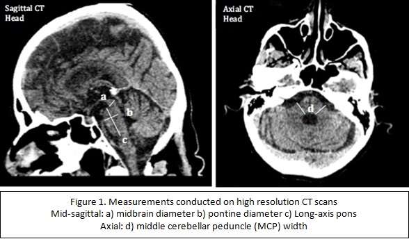
Methods: CT scans of patients with four diagnoses: PSP (n=13), MSA (n=6), Parkinson’s disease (PD: n=20; control), and essential tremor (ET: n=20; control) were retrospectively reviewed at a University teaching Hospital in South Wales, UK. CT scans (0.625mm isotropic voxel resolution) were reviewed using multi-planar reformat software. Two neuroradiology consultants, blinded to clinical information, carried out measurements: a) midbrain diameter (mid-sagittal), b) pontine diameter, c) long-axis pons and d) middle cerebellar peduncle (MCP) width to assess for structural changes linked to PSP and MSA [figure 1].
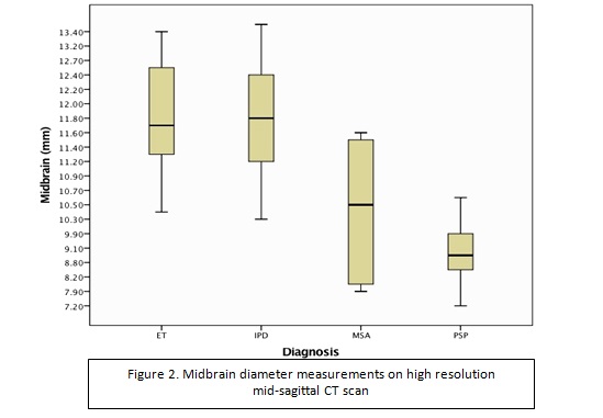
Results: Average midbrain diameter was significantly reduced in PSP (9.06mm ± 1.02mm) when compared with ET (11.89mm ± 0.91mm) and PD (11.79mm ± 0.84mm) [figure 2]. Receiver operating characteristic (ROC) analysis of midbrain measurements to differentiate between PSP and controls showed excellent sensitivity (Sn=92-100%) and specificity (Sp=80-98%) using cut-off values of 10.35mm-11.00mm. Similar analysis of midbrain measurements to differentiate between PSP and MSA gave a Sn of 77% and Sp of 66% at 10.00mm. Both MSA (15.40mm ± 2.43mm) and PSP (14.78mm ± 1.86mm) showed a significant reduction in MCP width when compared with controls (p<0.05). Intra and inter-rater analysis of midbrain measurements gave an intraclass correlation coefficient (ICC) > 0.7. Similar analysis of the pons gave an ICC of 0.55.
Conclusions: Our results show promise for the use of CT scans to aid diagnosis of PSP, especially when assessing midbrain atrophy. Loss of MCP width may be useful in the diagnosis of MSA, however, the Sn and Sp is lower and our study is limited by a small sample size (n=6). Overall, CT scans may not be a replacement of the gold-standard, MRI, but may be useful in guiding a differential diagnosis in patients with or without structural changes on CT.
To cite this abstract in AMA style:
S. Potluri, C. Gan, C. Thomas, B. Mohamed, S. Schwarz. Midbrain Diameter Measurements on High-Resolution Computer Tomography Scans to aid diagnosis of Progressive Supranuclear Palsy [abstract]. Mov Disord. 2018; 33 (suppl 2). https://www.mdsabstracts.org/abstract/midbrain-diameter-measurements-on-high-resolution-computer-tomography-scans-to-aid-diagnosis-of-progressive-supranuclear-palsy/. Accessed June 21, 2026.« Back to 2018 International Congress
MDS Abstracts - https://www.mdsabstracts.org/abstract/midbrain-diameter-measurements-on-high-resolution-computer-tomography-scans-to-aid-diagnosis-of-progressive-supranuclear-palsy/