Objective: To characterize white matter microstructural alterations in isolated REM sleep behavior disorder (iRBD) using both conventional diffusion tensor imaging and advanced Neurite Orientation Dispersion and Density Imaging (NODDI), and to examine associations between these changes and motor performance.
Background: iRBD is a parasomnia that reflects an evolving α-synucleinopathy disorder, providing an opportunity to study early pathological changes. While diffusion tensor imaging (DTI) studies have shown white matter changes in iRBD, NODDI may offer better biological specificity in characterizing microstructural alterations through measures of Neurite Density Index (NDI), Orientation Dispersion Index (ODI), and Free Water Fraction (FWF).
Method: We included 77 participants with polysomnography-confirmed iRBD from the North American Prodromal Synucleinopathy (NAPS) Consortium and 154 age- and sex-matched cognitively unimpaired controls from the Mayo Clinic Study of Aging [table1]. White matter microstructure was evaluated using standardized multi-shell diffusion on 3T MRI, quantifying DTI metrics (fractional anisotropy, FA; and mean diffusivity, MD) and NODDI parameters across white matter tracts defined by the “Eve” WM atlas [figure1]. Group differences were assessed using conditional logistic regression, with correlations to motor performance evaluated using the Purdue Pegboard test.
Results: Compared to controls, iRBD participants demonstrated widespread and bidirectional white matter changes across major white matter pathways [figure2]. While predominantly showing decreases, FA exhibited some increases, particularly in the corticospinal tract. MD showed a largely opposite pattern with predominantly increased values across tracts. NODDI metrics revealed complex bidirectional patterns: ODI was broadly increased across multiple tracts with focal decreases, while NDI showed a pattern of predominantly decreased values alongside localized increases. DTI and NODDI metrics showed extensive, moderate correlations with the Purdue Pegboard Test (T= 2.0, p < 0.05, [figure3]).
Conclusion: These results suggest that extensive white matter abnormalities occur early in prodromal α-synucleinopathies and highlight the value of advanced diffusion imaging techniques in characterizing iRBD and its potential prediction of phenoconversion to overt neurodegenerative disorders.
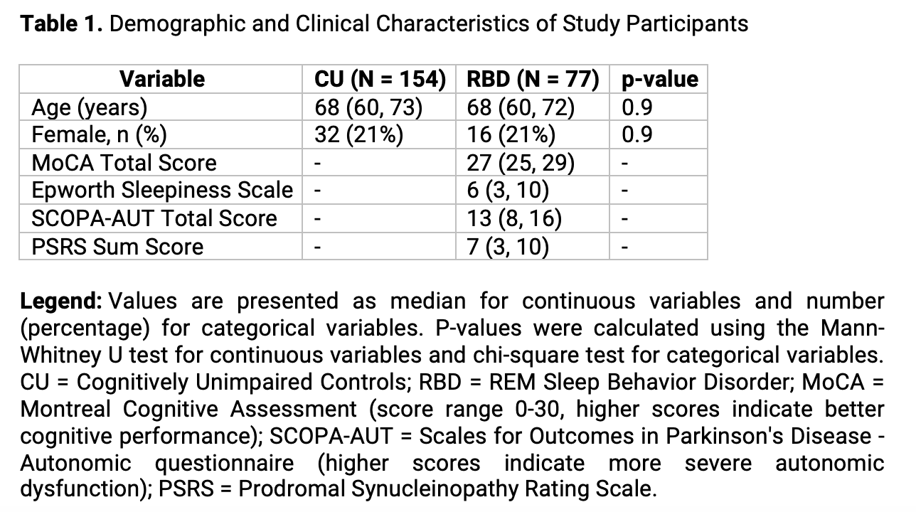
table1
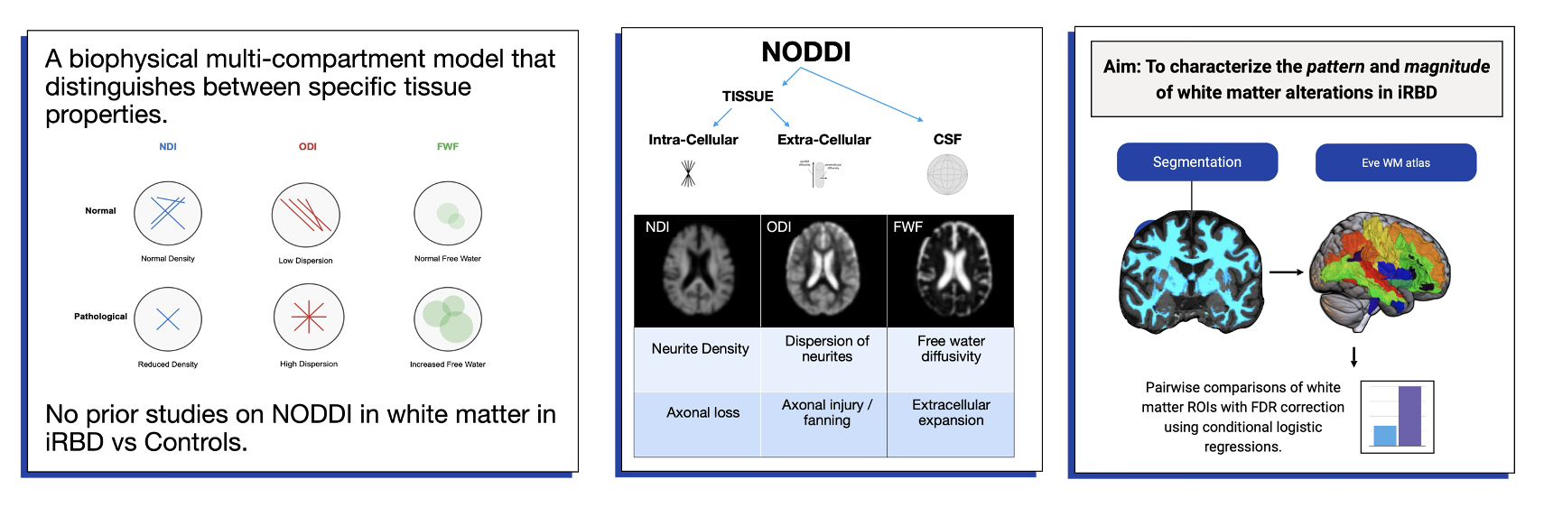
figure1
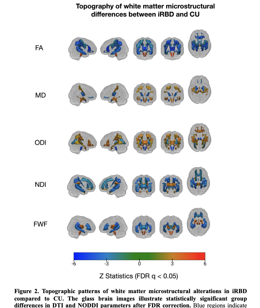
figure2
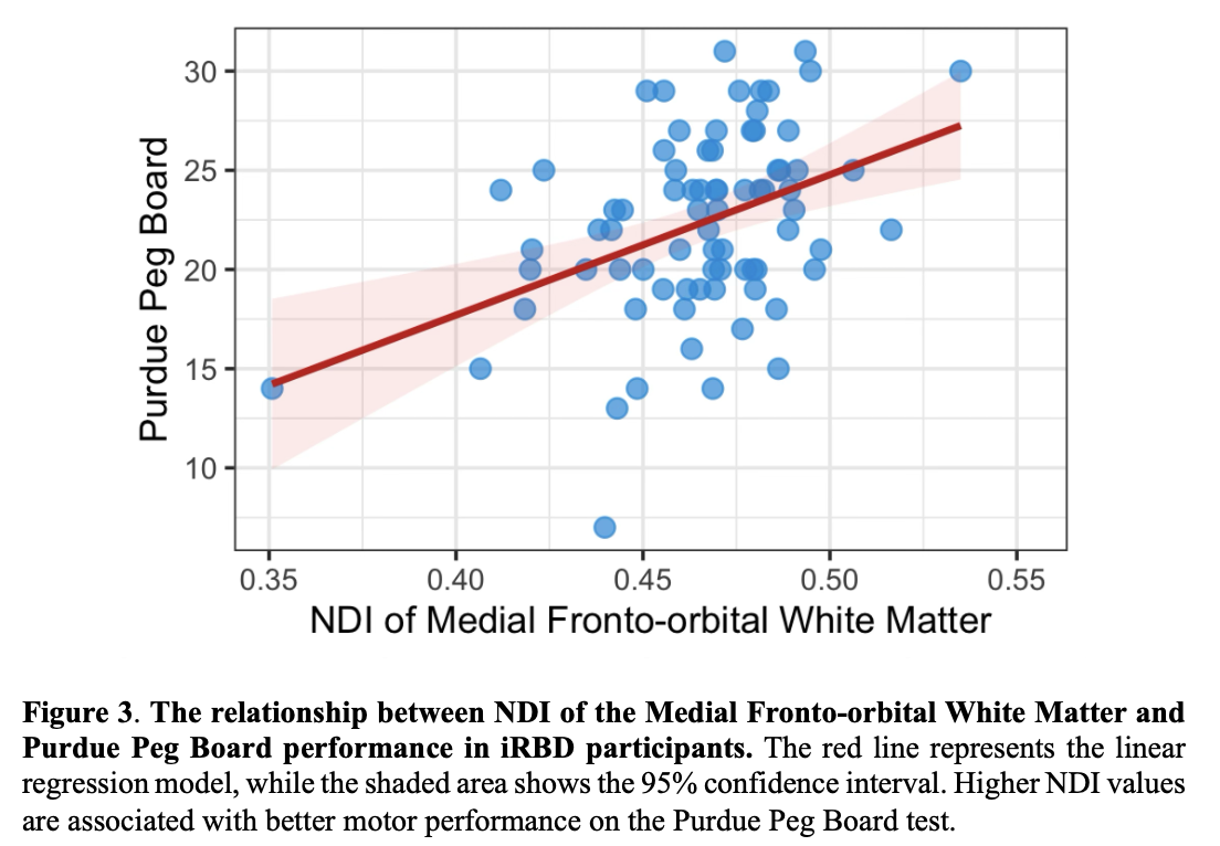
figure3
To cite this abstract in AMA style:
E. Mak, S. Przybelski, R. Reid, A. Fought, C. Schwarz, P. Vemuri, C. Jack JR., Y. Nie, H. Delacruz, A. Avidan, D. Bliwise, M. Campbell, S. Criswell, A. Davis, K. Duff, K. Ehgoetz Martens, J. Elliot, T. Ferman, J. Fields, L. Forsberg, J. Gagnon, Z. Gan-Or, M. Howell, M. Hu, X. Hu, D. Huddleston, P. Kotzbauer, J. Langley, M. Lim, J. Locke, V. Lowe, S. Mccarter, J. Mcleland, M. Miglis, E. Mignot, T. Miyagawa, L. Neilson, K. Nichols, A. Pelletier, O. Ross, C. Schenck, S. Wolfgang, E. ST. Louis, L. Trotti, A. Videnovic, C. Xiong, R. Postuma, Y. Ju, B. Boeve, K. Kantarci. Neurite Orientation Dispersion and Density Imaging revals reveals white matter microstructural changes in isolated REM sleep behavior disorder [abstract]. Mov Disord. 2025; 40 (suppl 1). https://www.mdsabstracts.org/abstract/neurite-orientation-dispersion-and-density-imaging-revals-reveals-white-matter-microstructural-changes-in-isolated-rem-sleep-behavior-disorder/. Accessed April 7, 2026.« Back to 2025 International Congress
MDS Abstracts - https://www.mdsabstracts.org/abstract/neurite-orientation-dispersion-and-density-imaging-revals-reveals-white-matter-microstructural-changes-in-isolated-rem-sleep-behavior-disorder/