Category: Rare Genetic and Metabolic Diseases
Objective: To evaluate the relationship between white matter (WM) changes and neurological impairment in Wilson disease (WD).
Background: Neuroimaging biomarkers derived from diffusion tensor imaging (DTI) have been proposed for WD, and changes in WM have been shown to correlate with neurological severity [1,2]. However, since evidence is limited and inconclusive [3], further validation is needed.
Method: 15 patients with WD (2 male; age: 41.5±11.5 years; disease duration: 218±100 months; on anti-copper treatment) were scored on the Unified Wilson’s Disease Rating Scale neurological subscale (UWDRS-N). 3T multi-shell diffusion MRI was acquired according to the Lifespan Human Connectome Project in Aging protocol and preprocessed with its minimal preprocessing pipeline [4]. DTI parameters, i.e., fractional anisotropy (FA) and mean diffusivity (MD), were computed using the Functional MRI of the Brain Software Library v.6.0.2 DTIFIT and voxelwise statistical analyses were carried out using RANDOMISE permutation testing (n=5000) in Tract-Based Spatial Statistics (TBSS, [5]). Significant clusters were identified by applying Threshold-Free Cluster Enhancement (p≤.05, family-wise error corrected) and labeled using the Johns Hopkins University WM atlas.
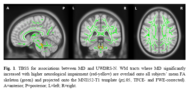
Results: TBSS revealed a significant positive association between MD and UWDRS-N scores (M=7.93±11.76) in the following WM tracts: bilateral superior cerebellar peduncles, right cerebellar peduncle, bilateral medial lemniscus, bilateral corticospinal tracts, and right genu of corpus callosum (see Fig. 1).
Conclusion: Our study found a significant increase of MD with higher neurological severity in WM tracts of WD patients, mainly affecting projection fibers near the brainstem and cerebellum, which may contribute to observed motor symptoms such as ataxia. These novel findings are consistent with previously demonstrated evidence of increased MD in the brain stem and midbrain in WD compared to controls [2,6]. Depending on the individual disease course, this may reflect acute inflammatory edemas or long-term neuronal loss and spongiform changes [6,7]. Although we did not find any significant FA changes related to UWDRS-N, the highly variable FA in WD across studies encourages us to focus on other markers of WM integrity like MD [3]. Overall, our findings suggest MD to be a promising neuroimaging biomarker even for residual neurological impairments in treated WD patients.
References: [1] Shribman, S., Bocchetta, M., Sudre, C. H., Acosta-Cabronero, J., Burrows, M., Cook, P., Thomas, D. L., Gillett, G. T., Tsochatzis, E. A., Bandmann, O., Rohrer, J. D., & Warner, T. T. (2022). Neuroimaging correlates of brain injury in Wilson’s disease: a multimodal, whole-brain MRI study. Brain, 145, 263-275. doi: 10.1093/brain/awab274.
[2] Jadav, R., Saini, J., Sinha, S., Bagepally, B., Rao, S., & Taly, A. B. (2013). Diffusion tensor imaging (DTI) and its clinical correlates in drug naive Wilson‘s disease. Metabolic Brain Disorders, 28, 455-462. doi: 10.1007/s11011-013-9407-1.
[3] Karimi, A., Mohammadi, S., Salehi, M. A., & Dager, S. R. (2022). Brain microstructural abnormalities in patients with Wilson’s disease: A systematic review of diffusion tensor imaging studies. Brain Imaging & Behavior, 16, 2809-2840. doi: 10.1007/s11682-022-00733-7.
[4] Glasser, M. F., Sotiropoulos, S. N., Wilson, J. A., Coalson, T. S., Fischl, B., Andersson, J. L., Xu, J., Jbabdi, S., Webster, M., Polimeni, J. R., Van Essen, D. C., Jenkinson, M., & Wu-Minn HCP Consortium. (2013). The minimal preprocessing pipelines for the Human Connectome Project. NeuroImage, 80, 105-124. doi: 10.1016/j.neuroimage.2013.04.127.
[5] Smith, S. M., Jenkinson, M., Johansen-Berg, H., Rueckert, D., Nichols, T.E., Mackay, C. E., Watkins, K. E., Ciccarelli, O., Cader, M. Z., Matthews, P. M., & Behrens, T. E. J. (2006). Tract-based spatial statistics: Voxelwise analysis of multi-subject diffusion data. NeuroImage, 31, 1487-1505. doi: 10.1016/j.neuroimage.2006.02.024.
[6] Wang, A., Wu, H., Xu, C., Tang, L., Lee, J., Wang, M., Jiang, M., Li, C., Lu, Q. & Zhang C. (2017). Study on lesion assessment of cerebello-thalamo-cortical network in Wilson’s disease with diffusion tensor imaging. Neural Plasticity, 2017, 7323121. doi: 10.1155/2017/7323121.
[7] Sener, R. N. (2003). Diffusion MRI findings in Wilson‘s disease. Computerized Medical Imaging and Graphics, 27, 17-21. doi: 10.1016/s0895-6111(02)00047-2.
To cite this abstract in AMA style:
A. Hausmann, S. Kannenberg, C. Hartmann, J. Caspers, C. Rubbert, A. Schnitzler. Neurological symptom severity in Wilson disease is associated with increased white matter mean diffusivity [abstract]. Mov Disord. 2023; 38 (suppl 1). https://www.mdsabstracts.org/abstract/neurological-symptom-severity-in-wilson-disease-is-associated-with-increased-white-matter-mean-diffusivity/. Accessed March 23, 2026.« Back to 2023 International Congress
MDS Abstracts - https://www.mdsabstracts.org/abstract/neurological-symptom-severity-in-wilson-disease-is-associated-with-increased-white-matter-mean-diffusivity/