Category: Technology
Objective: To assess the feasibility of a bright posterior limb of the internal capsule (pLIC) signal (PICS) in DiMANI images, as a reliable landmark for directly targeting the VIM in DBS procedures.
Background: Difficulties in direct visualization of thalamic subnuclei have led to inconsistent surgical outcomes1, 2 and repeat surgeries for patients undergoing DBS surgery for medication refractory tremors. Using the DiMANI contrast3, we present a new MRI landmark in the pLIC, referred to as PICS, that indicates the location of the Vim nucleus of the thalamus, enabling direct targeting approaches for surgical interventions.
Method: One healthy control and fifteen essential tremor (ET) patients were scanned at 7Tesla. To characterize the PICS fibers, two pLIC tractography schemes were conducted with cortical ROIs as seeds and the pLIC as a waypoint: i) gross motor cortical ROIs were extracted from the HMAT template4, ii) M1 homunculus was extracted from the Brainnetome data5. Finally, intra- and post-operative clinical data were merged for one ET DBS patient to show correspondence between the parcellation results and clinical observations.
Results: PICS was consistently identified across multiple MRI sequences, but more easily idendified in DiMANI sequences in all patients (Fig.1). Tractography analyses identified PICS to correlate with the distribution of motor fibers from the internal capsule (Fig.2A-C). For the M1 homunculus, two somatotopic clusters were observed: one including mostly trunk, lower and upper limbs; and another, more anteriorly, with head/face clustering with tongue/larynx (Fig.2D-F). Intra-operative stimulation at two different depths resulted in pLIC-specific side effect in the tongue/face. At those depths, measurements showed closer proximity of the DBS electrode to M1 clusters of head/face and tongue/larynx, validating the imaging findings (Fig.3).
Conclusion: PICS appears to be a reliable radiological marker comprising cortico-spinal tracts, in isolation from corticobulbar tracts fibers. It is consistently located lateral to the VIM, making it a potential landmark for targeting thalamic procedures. The parcellations of the pLIC using M1 homunculus could potentially inform lead or ablation location based on side effect profiles (e.g. head/face/tongue vs. trunk/limbs). Further studies are needed to correlate PICS with clinical outcomes and electrode locations in DBS procedures.
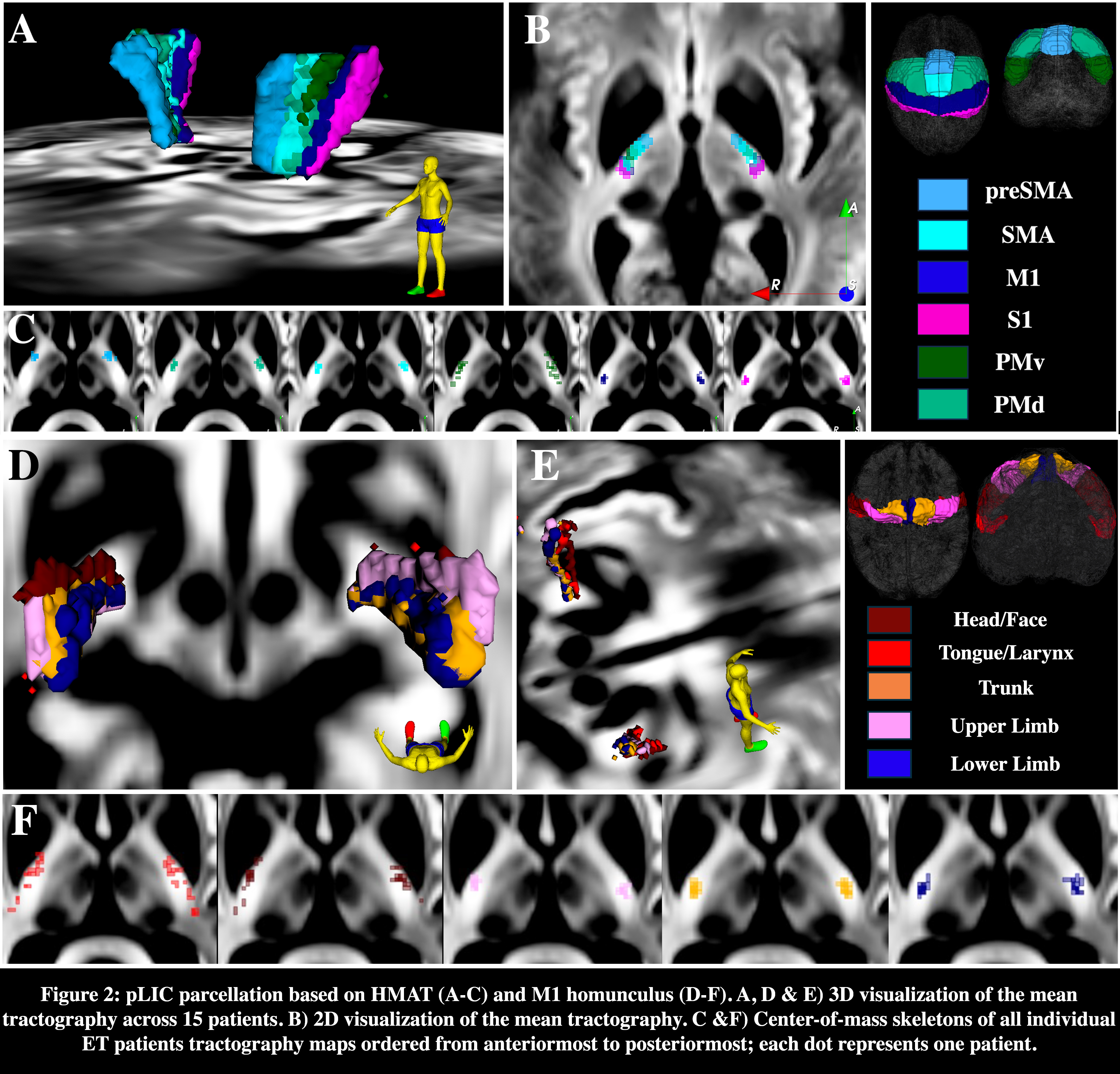
Fig.2
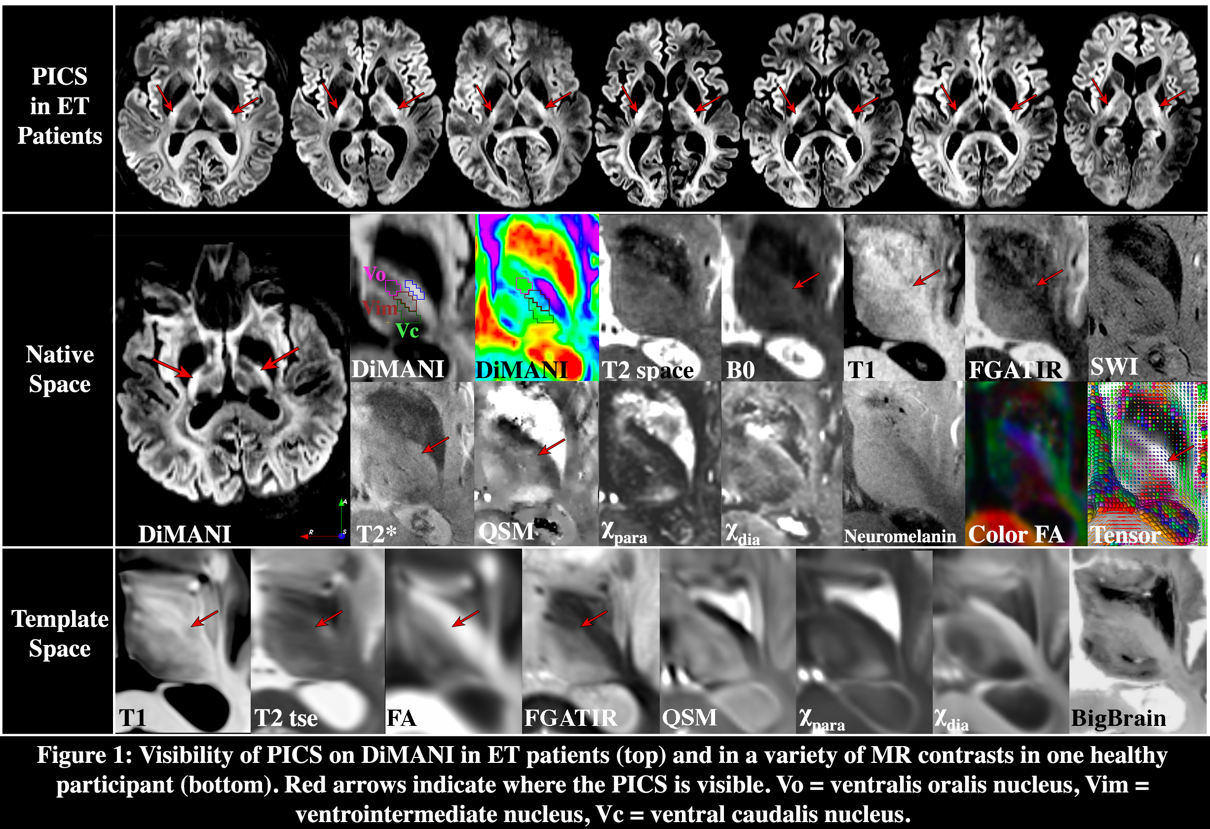
Fig.1

Fig.3
References: 1. Flora ED, Perera CL, Cameron AL, Maddern GJ. Deep brain stimulation for essential tremor: a systematic review. Mov Disord 2010;25(11):1550-1559.
2. Agrawal M, Garg K, Samala R, Rajan R, Naik V, Singh M. Outcome and Complications of MR Guided Focused Ultrasound for Essential Tremor: A Systematic Review and Meta-Analysis. Front Neurol 2021;12:654711.
3. Patriat R, Palnitkar T, Chandrasekaran J, et al. DiMANI: diffusion MRI for anatomical nuclei imaging-Application for the direct visualization of thalamic subnuclei. Front Hum Neurosci 2024;18:1324710.
4. Mayka MA, Corcos DM, Leurgans SE, Vaillancourt DE. Three-dimensional locations and boundaries of motor and premotor cortices as defined by functional brain imaging: a meta-analysis. Neuroimage 2006;31(4):1453-1474.
5. Fan L, Li H, Zhuo J, et al. The Human Brainnetome Atlas: A New Brain Atlas Based on Connectional Architecture. Cereb Cortex 2016;26(8):3508-3526.
To cite this abstract in AMA style:
R. Patriat, J. Chandrasekaran, K. Sretavan, H. Braun, S. Brenny, Y. Seddighi, J. Aman, M. Hill, J. Vitek, N. Harel, L. Almeida. “PICS”: a Novel Radiological Landmark for Thalamic DBS Interventions [abstract]. Mov Disord. 2025; 40 (suppl 1). https://www.mdsabstracts.org/abstract/pics-a-novel-radiological-landmark-for-thalamic-dbs-interventions/. Accessed April 13, 2026.« Back to 2025 International Congress
MDS Abstracts - https://www.mdsabstracts.org/abstract/pics-a-novel-radiological-landmark-for-thalamic-dbs-interventions/