Session Information
Date: Monday, October 8, 2018
Session Title: Parkinson's Disease: Neuroimaging And Neurophysiology
Session Time: 1:15pm-2:45pm
Location: Hall 3FG
Objective: To perform an exhaustive comparison of brain networks identified through functional MRI (fMRI) in a small cohort of patients with idiopathic Parkinson’s disease (IPD) at an early stage, compared to healthy controls (HC), and to discuss their potential role in disease pathogenesis and clinical phenotype.
Background: Brain network connectivity investigated in IPD by fMRI has yielded highly variable results with overlapping disease-driven and compensatory changes, especially for early disease stages, when only subtle alterations are detectable. However, exhaustive an unbiased studies of resting state networks (RSNs), beyond those already known to be involved in IPD such as the basal ganglia (BGN), the default mode (DMN) and the sensory-motor networks, have rarely being carried out.
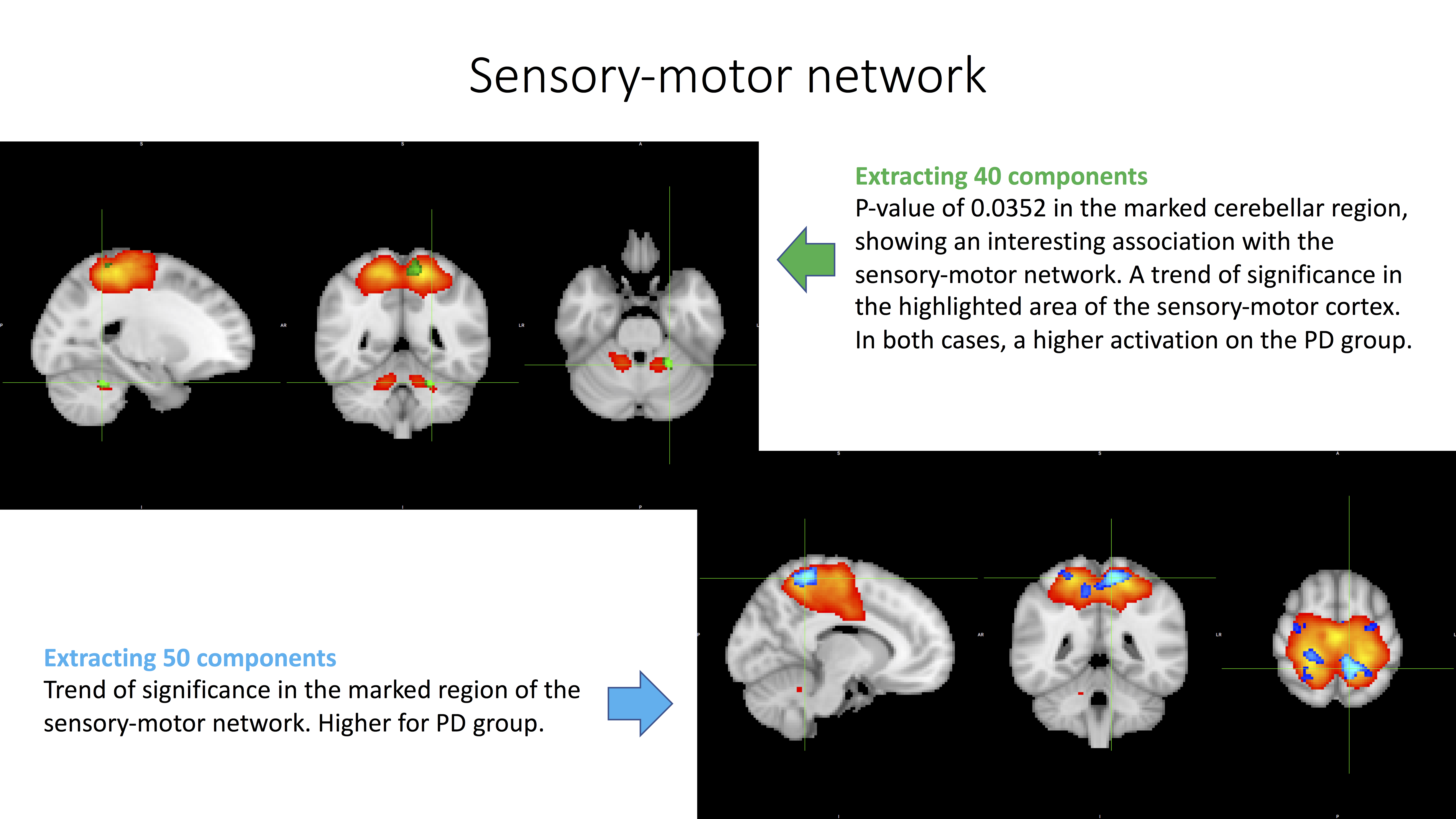
Methods: RS-fMRI was performed in 20 early IPD patients (age: 57.6 ± 9.6 years, disease duration 4.35 ± 2.2 years) and 7 HC (age: 58 ± 5.41years). The IPD patients were off dopaminergic medications for ≥12 hours. The brain networks were extracted from the fMRI sequences by using independent component (IC) analysis. The data was cleaned by removing artefactual networks, and used to obtain average ICs. We identified clusters with significant differences between both groups by performing group-wise statistical comparisons. All steps were run with the FSL tools Melodic, Fix and Randomise.
Results: After running analyses for both 40 and 50 average ICs, standard RSNs, including the DMN, the BGN, various visual networks, etc., were consistently identified. The comparison in the 40 ICs analysis showed higher activation in IPD patients for the RSNs: sensory-motor, cerebellar, and parts of high-order cognition (p-values < 0.05). And the comparison for the 50 ICs analysis also showed higher activation in IPD for the precuneus, DMN, and primary visual RSNs (p-values < 0.05). For the sensory-motor, DMN and precuneus RSNs, the significant differences for one analysis were confirmed by a trend for significance on the second [figure1]. FSL tool Randomise controls for family-wise error and enhances clusters of voxels.
Conclusions: This pilot study suggests that extensive fMRI exploration, surprisingly, identifies disparate network activations in early IPD patients, beyond the normally reported RSNs, such as the BGN and DMN. While similar results have been reported for the cerebellar network, higher activation of the primary visual network may pinpoint to the risk of visual symptoms in IPD patients. Due to the small sample size, robustness of the obtained results warrants confirmation in independent and larger cohorts. Besides, the impact of compensatory network activations on clinical phenotype and disease trajectory needs further investigation.
To cite this abstract in AMA style:
L. Salamanca, E. Glaab, R. Balling, G. Dooms, N. Diederich. Resting state fMRI points to disparate brain network activations in early Parkinson’s disease [abstract]. Mov Disord. 2018; 33 (suppl 2). https://www.mdsabstracts.org/abstract/resting-state-fmri-points-to-disparate-brain-network-activations-in-early-parkinsons-disease/. Accessed May 20, 2026.« Back to 2018 International Congress
MDS Abstracts - https://www.mdsabstracts.org/abstract/resting-state-fmri-points-to-disparate-brain-network-activations-in-early-parkinsons-disease/