Objective: To identify distinct pallidal local field potential (LFP) signatures characterizing status dystonicus (SD) from non-SD dystonia.
Background: SD is a poorly understood neurological emergency requiring urgent interventions, including globus pallidus interna (GPi) deep brain stimulation (DBS). Sensing-capable DBS electrodes provide an opportunity to study SD pathophysiology. Despite increasing use of pallidal DBS, oscillatory & non-oscillatory electrophysiological signatures of SD remain unknown. We sought to identify intracranial neural biomarkers of SD from a rare cohort of children, critical since children at risk often have chronic baseline dystonia difficult to differentiate from SD.
Method: Ten patients (age: 7.8+/-3.6) with SD (Hospital for Sick Children, Canada) were implanted with Medtronic Percept™ GPi DBS [figure1](A). LFPs were recorded longitudinally during SD, recovery and relapse (range: 11-1155 days) and Power Spectral Densities (PSDs) calculated (Welch-method). Analysis of oscillatory and aperiodic activity was performed [figure2](A) (fitting-oscillations/one-over-f). Band-limited power (Theta: 3-7Hz; Alpha: 7-12.5Hz, Beta: 12.5-30Hz) was calculated. Bursting activity within +/-3 Hz around the beta peak frequency was computed [figure3](A). Generalized linear mixed effects models assessed relations between LFP metrics and clinical scales, including Burke‐Fahn‐Marsden Dystonia Rating Scale (BFMDRS) and Pediatric Quality of Life Score (PedsQL). Circadian LFP dynamics were assessed in one participant with narrowband beta-band recordings over months.
Results: During SD, periodic activity in the beta-band increased across all patients and recordings [figure1](B), with increased periodic peak power [figure2](B) (p=0.007) and beta-band burst amplitude [figure3](B&C) (p<0.001). Relapsed SD was characterized by return of excessive beta signatures [figure1](C). Beta-specific LFP power alone was significantly associated with worse quality-of-life scores [figure4] (PedsQL, p=0.03, R2=0.695). Circadian beta-band periodicity was present [figure5](A), with significantly increased power across daytime (p<0.001) and nighttime (p<0.001) during SD [figure5](B).
Conclusion: SD is a distinct state with important implications for dystonia pathophysiology, tracking dystonia-states from intracranial activity and potential adaptive DBS treatments.
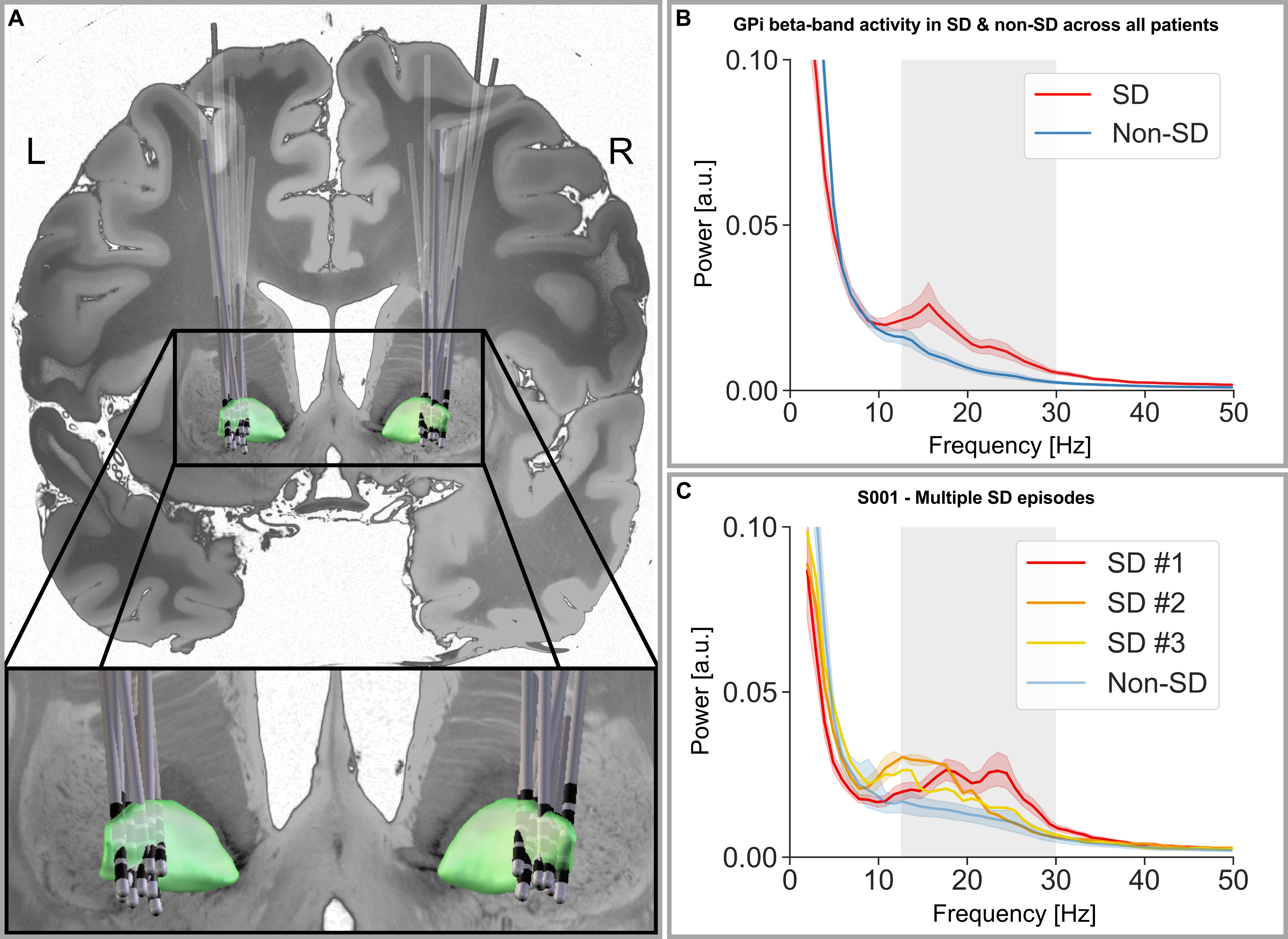
figure1
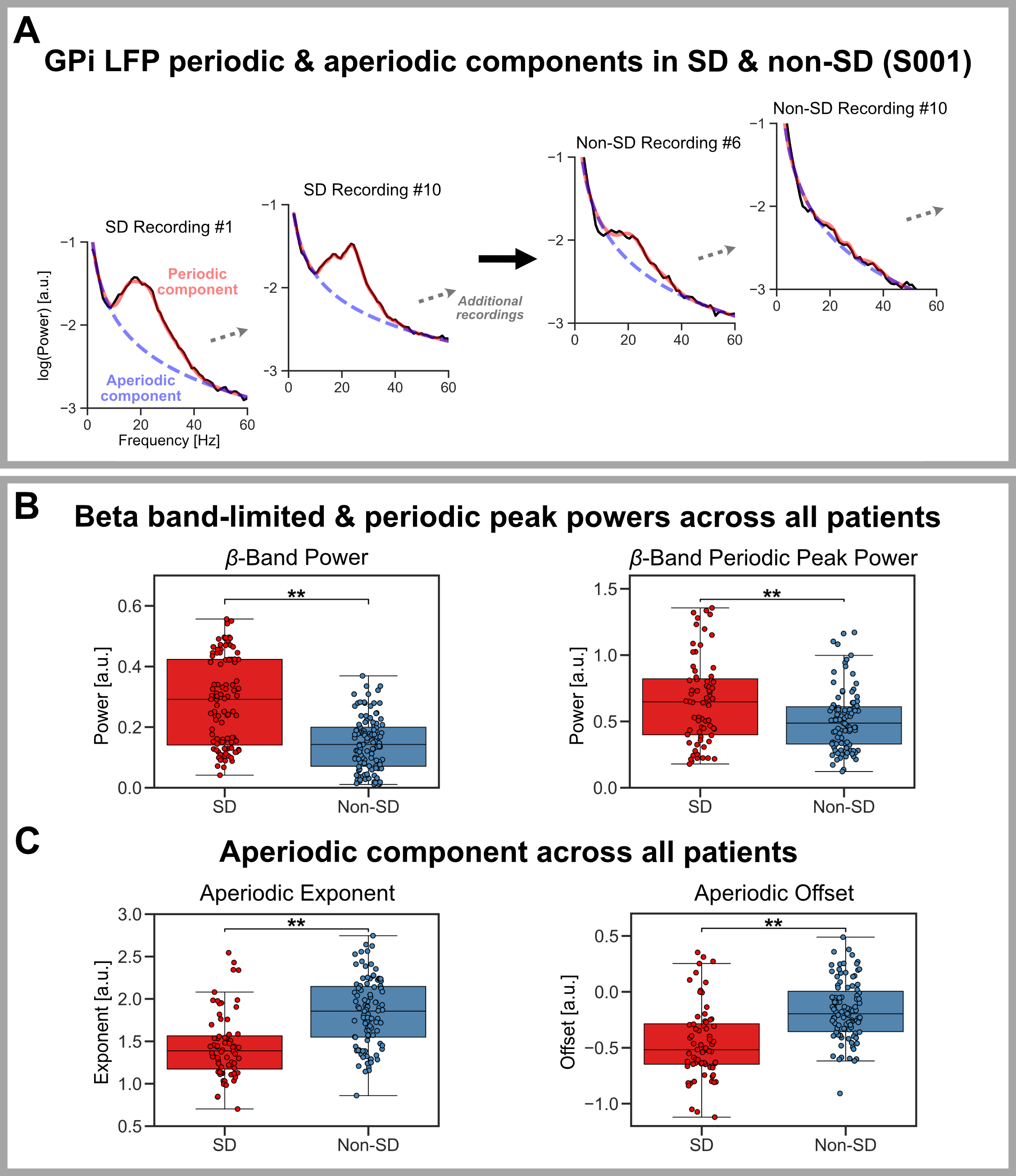
figure2
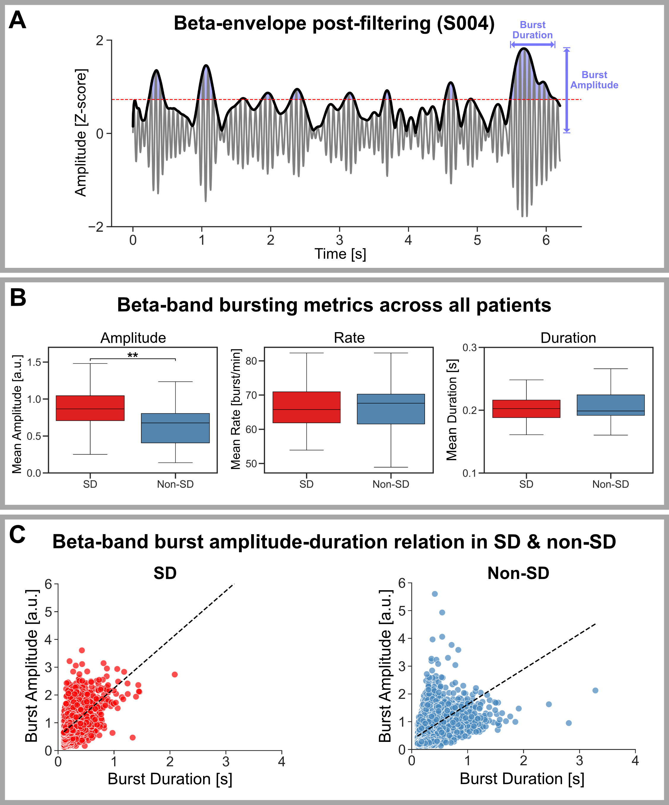
figure3
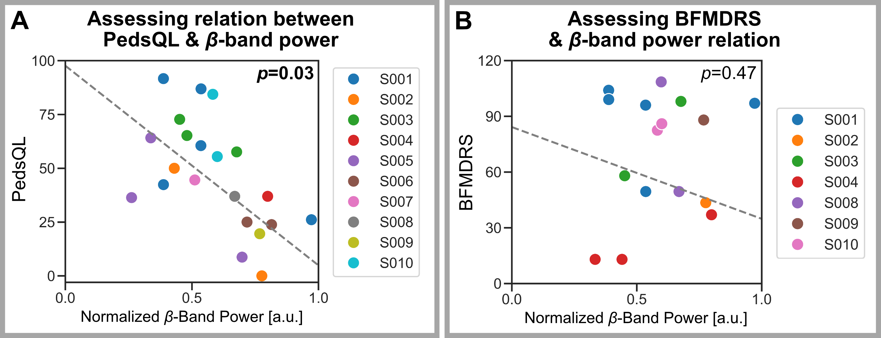
figure4
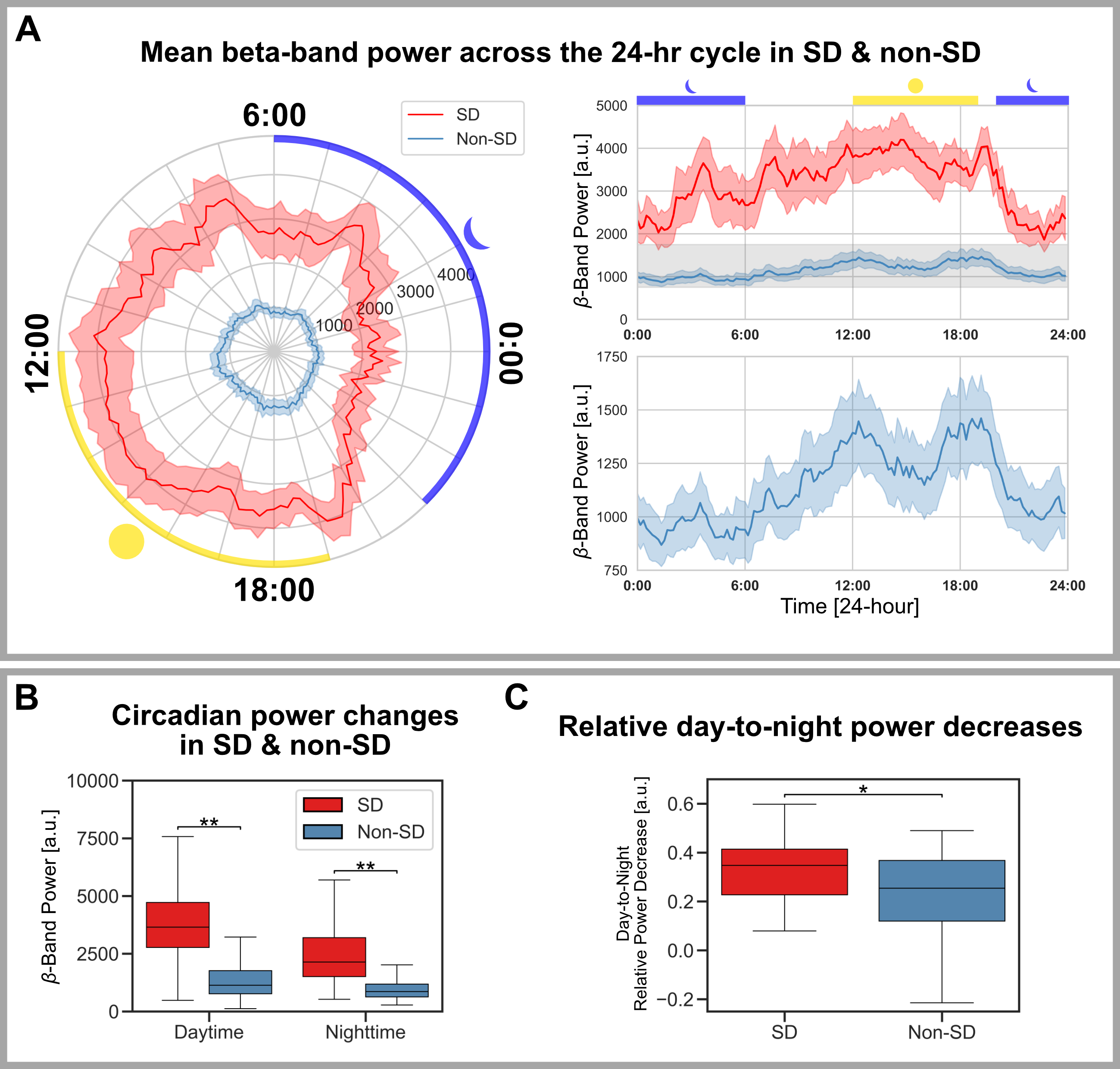
figure5
References: 1. Albanese, A. et al. Phenomenology and classification of dystonia: A consensus update. Movement Disorders 28, 863–873 (2013).
2. Goswami, J. N., Roy, S. & Patnaik, S. K. Pediatric Dystonic Storm: A Hospital-Based Study. Neurology: Clinical Practice 11, e645 (2021).
3. Saini, A. G. et al. Status Dystonicus in Children: A Cross-Sectional Study and Review of Literature. J Child Neurol 37, 441–450 (2022).
4. Meijer, I. A. & Fasano, A. Status Dystonicus. in Movement Disorder Emergencies: Diagnosis and Treatment (ed. Frucht, S. J.) 183–199 (Springer International Publishing, Cham, 2022). doi:10.1007/978-3-030-75898-1_10.
5. Vogt, L. M. et al. Recommendations for the Management of Initial and Refractory Pediatric Status Dystonicus. Movement Disorders 39, 1435–1445 (2024).
6. Ruiz-Lopez, M. & Fasano, A. Rethinking status dystonicus. Mov Disord 32, 1667–1676 (2017).
7. Allen, N. M., Lin, J.-P., Lynch, T. & King, M. D. Status dystonicus: a practice guide. Developmental Medicine & Child Neurology 56, 105–112 (2014).
8. Vogt, L. M. et al. Deep Brain Stimulation for Refractory Status Dystonicus in Children: Multicenter Case Series and Systematic Review. Annals of Neurology 95, 156–173 (2024).
9. Benato, A. et al. Long-term effect of subthalamic and pallidal deep brain stimulation for status dystonicus in children with methylmalonic acidemia and GNAO1 mutation. J Neural Transm (Vienna) 126, 739–757 (2019).
10. Ben-Haim, S. et al. Deep Brain Stimulation for Status Dystonicus: A Case Series and Review of the Literature. Stereotactic and Functional Neurosurgery 94, 207–215 (2016).
11. Lobato-Polo, J. et al. Deep Brain Stimulation Surgery for Status Dystonicus: A Single-Center Experience and Literature Review. World Neurosurgery 114, e992–e1001 (2018).
12. Simonyan, K., Cho, H., Hamzehei Sichani, A., Rubien-Thomas, E. & Hallett, M. The direct basal ganglia pathway is hyperfunctional in focal dystonia. Brain 140, 3179–3190 (2017).
13. Tisch, S. & Limousin, P. Neurophysiological insights in dystonia and its response to deep brain stimulation treatment. Exp Brain Res 238, 1645–1657 (2020).
14. Quartarone, A. & Hallett, M. Emerging concepts in the physiological basis of dystonia. Movement Disorders 28, 958–967 (2013).
15. Lofredi, R. et al. Striato-pallidal oscillatory connectivity correlates with symptom severity in dystonia patients. Nat Commun 15, 8475 (2024).
16. Scheller, U. et al. Pallidal low-frequency activity in dystonia after cessation of long-term deep brain stimulation. Movement Disorders 34, 1734–1739 (2019).
17. Neumann, W.-J. et al. A localized pallidal physiomarker in cervical dystonia. Annals of Neurology 82, 912–924 (2017).
18. Neumann, W.-J. et al. Enhanced low-frequency oscillatory activity of the subthalamic nucleus in a patient with dystonia. Movement Disorders 27, 1063–1066 (2012).
19. Geng, X. et al. Comparison of oscillatory activity in subthalamic nucleus in Parkinson’s disease and dystonia. Neurobiol Dis 98, 100–107 (2017).
20. Lofredi, R. et al. Pallidal beta bursts in Parkinson’s disease and dystonia. Movement Disorders 34, 420–424 (2019).
21. Little, S. & Brown, P. What brain signals are suitable for feedback control of deep brain stimulation in Parkinson’s disease? Annals of the New York Academy of Sciences 1265, 9–24 (2012).
22. Neumann, W.-J. et al. Subthalamic synchronized oscillatory activity correlates with motor impairment in patients with Parkinson’s disease. Movement Disorders 31, 1748–1751 (2016).
23. Tinkhauser, G. et al. Beta burst coupling across the motor circuit in Parkinson’s disease. Neurobiol Dis 117, 217–225 (2018).
24. Piña-Fuentes, D. et al. The characteristics of pallidal low-frequency and beta bursts could help implementing adaptive brain stimulation in the parkinsonian and dystonic internal globus pallidus. Neurobiol Dis 121, 47–57 (2019).
25. Tinkhauser, G. et al. Beta burst dynamics in Parkinson’s disease OFF and ON dopaminergic medication. Brain 140, 2968–2981 (2017).
26. Yu, Y. et al. Parkinsonism Alters Beta Burst Dynamics across the Basal Ganglia–Motor Cortical Network. The Journal of Neuroscience 41, 2274 (2021).
27. Lofredi, R. et al. Subthalamic beta bursts correlate with dopamine-dependent motor symptoms in 106 Parkinson’s patients. npj Parkinsons Dis. 9, 1–9 (2023).
28. Darmani, G. et al. Long-Term Recording of Subthalamic Aperiodic Activities and Beta Bursts in Parkinson’s Disease. Movement Disorders 38, 232–243 (2023).
29. Gao, R., Peterson, E. J. & Voytek, B. Inferring synaptic excitation/inhibition balance from field potentials. NeuroImage 158, 70–78 (2017).
30. Manning, J. R., Jacobs, J., Fried, I. & Kahana, M. J. Broadband Shifts in Local Field Potential Power Spectra Are Correlated with Single-Neuron Spiking in Humans. J. Neurosci. 29, 13613–13620 (2009).
31. Voytek, B. & Knight, R. T. Dynamic Network Communication as a Unifying Neural Basis for Cognition, Development, Aging, and Disease. Biological Psychiatry 77, 1089–1097 (2015).
32. Wiest, C. et al. Subthalamic Nucleus Stimulation–Induced Local Field Potential Changes in Dystonia. Movement Disorders 38, 423–434 (2023).
33. Iodice, A. & Pisani, F. Status dystonicus: management and prevention in children at high risk. Acta Biomed 90, 207–212 (2019).
34. Yin, Z. et al. Pallidal activities during sleep and sleep decoding in dystonia, Huntington’s, and Parkinson’s disease. Neurobiology of Disease 182, 106143 (2023).
35. Yin, Z. et al. Generalized sleep decoding with basal ganglia signals in multiple movement disorders. npj Digit. Med. 7, 1–13 (2024).
36. Anjum, M. F. et al. Multi-night cortico-basal recordings reveal mechanisms of NREM slow-wave suppression and spontaneous awakenings in Parkinson’s disease. Nat Commun 15, 1793 (2024).
37. Cagle, J. N. et al. Chronic intracranial recordings in the globus pallidus reveal circadian rhythms in Parkinson’s disease. Nat Commun 15, 4602 (2024).
38. Baud, M. O. et al. Multi-day rhythms modulate seizure risk in epilepsy. Nat Commun 9, 88 (2018).
39. Provenza, N. R. et al. Disruption of neural periodicity predicts clinical response after deep brain stimulation for obsessive-compulsive disorder. Nat Med 30, 3004–3014 (2024).
40. Neumann, W.-J., Gilron, R., Little, S. & Tinkhauser, G. Adaptive Deep Brain Stimulation: From Experimental Evidence Toward Practical Implementation. Movement Disorders 38, 937–948 (2023).
41. Balachandar, A., Phokaewvarangkul, O. & Fasano, A. Closed-loop systems for deep brain stimulation to treat neuropsychiatric disorders. Expert Review of Medical Devices 0, 1–12.
42. Phokaewvarangkul, O., Balachandar, A. & Fasano, A. Chapter 17 – Closed-loop systems. in Handbook of Digital Technologies in Movement Disorders (eds. Bhidayasiri, R. & Maetzler, W.) 269–284 (Academic Press, 2024). doi:10.1016/B978-0-323-99494-1.00002-2.
43. Gilron, R. et al. Sleep-Aware Adaptive Deep Brain Stimulation Control: Chronic Use at Home With Dual Independent Linear Discriminate Detectors. Front. Neurosci. 15, (2021).
44. Smyth, C. et al. Adaptive Deep Brain Stimulation for sleep stage targeting in Parkinson’s disease. Brain Stimulation: Basic, Translational, and Clinical Research in Neuromodulation 16, 1292–1296 (2023).
45. Yan, H. et al. The Child & Youth CompreHensIve Longitudinal Database for Deep Brain Stimulation (CHILD-DBS). Childs Nerv Syst 37, 607–615 (2021).
46. Horn, A. et al. Lead-DBS v2: Towards a comprehensive pipeline for deep brain stimulation imaging. Neuroimage 184, 293–316 (2019).
47. Yokochi, F. et al. Resting-State Pallidal-Cortical Oscillatory Couplings in Patients With Predominant Phasic and Tonic Dystonia. Front. Neurol. 9, (2018).
48. Fasano, A. et al. Status dystonicus: Predictors of outcome and progression patterns of underlying disease. Movement Disorders 27, 783–788 (2012).
49. Burke, R. E. et al. Validity and reliability of a rating scale for the primary torsion dystonias. Neurology 35, 73–77 (1985).
50. Pintér, D., Janszky, J. & Kovács, N. Minimal Clinically Important Differences for Burke-Fahn-Marsden Dystonia Rating Scale and 36-Item Short-Form Health Survey. Movement Disorders 35, 1218–1223 (2020).
51. Donoghue, T. et al. Parameterizing neural power spectra into periodic and aperiodic components. Nat Neurosci 23, 1655–1665 (2020).
52. Desai, A. D. et al. Validity and Responsiveness of the Pediatric Quality of Life Inventory (PedsQL) 4.0 Generic Core Scales in the Pediatric Inpatient Setting. JAMA Pediatrics 168, 1114–1121 (2014).
53. Varni, J. W., Seid, M. & Kurtin, P. S. PedsQL 4.0: reliability and validity of the Pediatric Quality of Life Inventory version 4.0 generic core scales in healthy and patient populations. Med Care 39, 800–812 (2001).
54. Varni, J. W., Limbers, C. A. & Burwinkle, T. M. Impaired health-related quality of life in children and adolescents with chronic conditions: a comparative analysis of 10 disease clusters and 33 disease categories/severities utilizing the PedsQLTM 4.0 Generic Core Scales. Health and Quality of Life Outcomes 5, 43 (2007).
55. Lofredi, R. et al. Pallidal Beta Activity Is Linked to Stimulation-Induced Slowness in Dystonia. Movement Disorders 38, 894–899 (2023).
56. Alamri, A. et al. Deep Brain Stimulation of the Globus Pallidus Internus in a Child with Refractory Dystonia due to L2-Hydroxyglutaric Aciduria. Stereotactic and Functional Neurosurgery 102, 209–216 (2024).
57. Fasano, A. et al. Local Field Potential-Based Programming: A Proof-of-Concept Pilot Study. Neuromodulation: Technology at the Neural Interface n/a, (2021).
58. Tinkhauser, G. et al. The modulatory effect of adaptive deep brain stimulation on beta bursts in Parkinson’s disease. Brain 140, 1053–1067 (2017).
59. Brittain, J.-S. & Brown, P. Oscillations and the basal ganglia: Motor control and beyond. NeuroImage 85, 637–647 (2014).
60. Weinberger, M. & Dostrovsky, J. O. A basis for the pathological oscillations in basal ganglia: the crucial role of dopamine. NeuroReport 22, 151 (2011).
61. Wang, D. D. et al. Pallidal Deep-Brain Stimulation Disrupts Pallidal Beta Oscillations and Coherence with Primary Motor Cortex in Parkinson’s Disease. J. Neurosci. 38, 4556–4568 (2018).
62. Mink, J. W. Special concerns in defining, studying, and treating dystonia in children. Movement Disorders 28, 921–925 (2013).
63. Gorodetsky, C. & Fasano, A. Approach to the Treatment of Pediatric Dystonia. Dystonia 1, 10287 (2022).
64. Gimeno, H. et al. Evaluation of functional goal outcomes using the Canadian Occupational Performance Measure (COPM) following Deep Brain Stimulation (DBS) in childhood dystonia. European Journal of Paediatric Neurology 18, 308–316 (2014).
65. Gimeno, H., Gordon, A., Tustin, K. & Lin, J.-P. Functional priorities in daily life for children and young people with dystonic movement disorders and their families. European Journal of Paediatric Neurology 17, 161–168 (2013).
66. Gimeno, H., Tustin, K., Selway, R. & Lin, J.-P. Beyond the Burke–Fahn–Marsden Dystonia Rating Scale: Deep brain stimulation in childhood secondary dystonia. European Journal of Paediatric Neurology 16, 501–508 (2012).
67. Kuiper, M. J. et al. The Burke-Fahn-Marsden Dystonia Rating Scale is Age-Dependent in Healthy Children. Movement Disorders Clinical Practice 3, 580–586 (2016).
68. Wiest, C. et al. The aperiodic exponent of subthalamic field potentials reflects excitation/inhibition balance in Parkinsonism. eLife 12, e82467 (2023).
69. Gilron, R. et al. Long-term wireless streaming of neural recordings for circuit discovery and adaptive stimulation in individuals with Parkinson’s disease. Nature Biotechnology 39, 1078–1085 (2021).
70. Pedregosa, F. et al. Scikit-learn: Machine Learning in Python. Journal of Machine Learning Research 12, 2825–2830 (2011).
To cite this abstract in AMA style:
A. Balachandar, L. Vogt, K. Mithani, S. Coleman, M. Ebden, A. Leblanc-Miller, S. Breitbart, A. Fasano, C. Gorodetsky, G. Ibrahim. Status Dystonicus is a Distinct State Characterized by Pallidal Beta-band Activity [abstract]. Mov Disord. 2025; 40 (suppl 1). https://www.mdsabstracts.org/abstract/status-dystonicus-is-a-distinct-state-characterized-by-pallidal-beta-band-activity/. Accessed April 6, 2026.« Back to 2025 International Congress
MDS Abstracts - https://www.mdsabstracts.org/abstract/status-dystonicus-is-a-distinct-state-characterized-by-pallidal-beta-band-activity/