Category: Dystonia: Pathophysiology, Imaging
Objective: _
Background: Typing on a keyboard requires complex collaboration between visuospatial procedural memory, language and motor function [1,2]. The impaired ability to type, independent of motor deficits, apraxia or aphasia has been coined “dystypia” [3]. Dystypia can be disruptive, given the ubiquity of technology in daily life. In light of recent reports of lost ability to swim after deep brain stimulation (DBS), we encountered a peculiar case of dystypia as a potential side effect of DBS therapy.
Method: Case report: A 68-year-old female with a history of blepharospasm, oromandibular and cervical segmental dystonia underwent bilateral pallidal DBS after her response to botulinum toxin therapy waned. Shortly after implantation, she reports “forgetting how to type on the computer”, while at baseline she was proficient in typing from prior office work. Following DBS, she discovered she no longer “remembered” how to type fluidly and had to “hunt and peck” for letters on the keyboard, persisting during stimulation even at two-year follow-up.
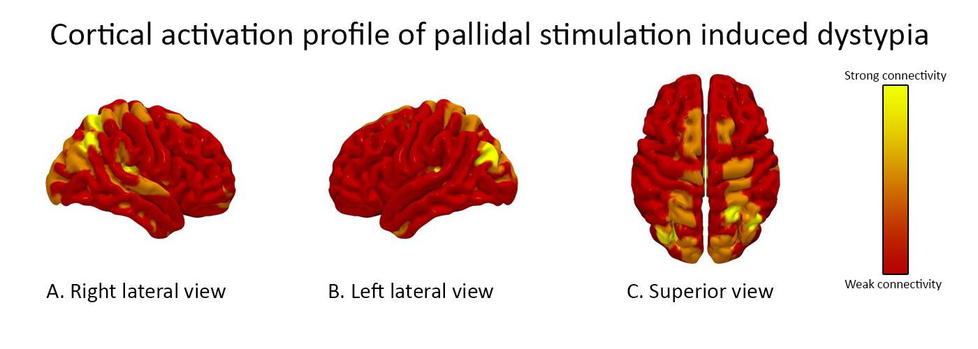
Post-operative lead reconstruction was performed using Lead-DBS [4,5]. Volume of tissue activation (VTA) modeling was then combined with whole brain tractography [6,7]. The resulting fiber tract activation pattern is shown [figure1]. Cortical mapping revealed strong modulation of the right supramarginal gyrus, left calcarine fissure and left cuneus. There was also activation of bilateral supplemental motor areas and superior parietal gyri.
Results: Discussion: Several case reports have described dystypia in the absence of motor weakness, apraxia and aphasia [1–3,8,9]. Typically these occurred in the setting of acute strokes. One case report described a patient with dystypia following bilateral subthalamic nucleus (STN) DBS [10]. Post-operative imaging discovered prominent vasogenic edema in the frontal lobe surrounding the left STN lead. A recent literature review investigating the shared lesion topography of dystypia cases proposed involvement of the superior longitudinal fasciculus (SLF) [8]. The SLF has been characterized to involve the superior parietal lobe, angular gyrus, supramarginal gyrus, and arcuate fasiculus [11]. Our patient’s connectivity pattern also suggests involvement of the SLF.
Conclusion: Dystypia is a rare side effect of DBS therapy and may be related to SLF involvement.
References: [1] Ryu D-W, Kim J-S, Yang D-W, Kim Y-I, Lee K-S. Dystypia Without Aphasia Associated With Visuospatial Memory Impairment in a Patient With Acute Stroke: Alzheimer Dis Assoc Disord 2012;26:285–8. https://doi.org/10.1097/WAD.0b013e318231e614. [2] Cook FAB, Makin SDJ, Wardlaw J, Dennis MS. Dystypia in acute stroke not attributable to aphasia or neglect. Case Rep 2013;2013:bcr2013200257–bcr2013200257. https://doi.org/10.1136/bcr-2013-200257. [3] Otsuki M, Soma Y, Arihiro S, Watanabe Y, Moriwaki H, Naritomi H. Dystypia: Isolated Typing Impairment without Aphasia, Apraxia or Visuospatial Impairment. Eur Neurol 2002;47:136–40. https://doi.org/10.1159/000047971. [4] Horn A, Kühn AA. Lead-DBS: A toolbox for deep brain stimulation electrode localizations and visualizations. NeuroImage 2015;107:127–35. https://doi.org/10.1016/j.neuroimage.2014.12.002. [5] Horn A, Li N, Dembek TA, Kappel A, Boulay C, Ewert S, et al. Lead-DBS v2: Towards a comprehensive pipeline for deep brain stimulation imaging. NeuroImage 2019;184:293–316. https://doi.org/10.1016/j.neuroimage.2018.08.068. [6] Yeh F-C, Verstynen TD, Wang Y, Fernández-Miranda JC, Tseng W-YI. Deterministic Diffusion Fiber Tracking Improved by Quantitative Anisotropy. PLOS ONE 2013;8:e80713. https://doi.org/10.1371/journal.pone.0080713. [7] Yeh F-C, Panesar S, Fernandes D, Meola A, Yoshino M, Fernandez-Miranda JC, et al. Population-averaged atlas of the macroscale human structural connectome and its network topology. NeuroImage 2018;178:57–68. https://doi.org/10.1016/j.neuroimage.2018.05.027. [8] Sharma AK, Fridman S, Gleichgerrcht E, Sposato LA. Dystextia and dystypia as modern stroke symptoms: A case series and literature review. Clin Neurol Neurosurg 2019;180:25–7. https://doi.org/10.1016/j.clineuro.2019.02.001. [9] Thomas NWD, Mestre TA. Impact of New Technologies in a Stroke Presentation: A Case of Dystextia and Dystypia. Can J Neurol Sci J Can Sci Neurol 2017;44:458–60. https://doi.org/10.1017/cjn.2016.431. [10] Lee S-H, Jeon K, Son B, Kim J-S. Isolated dystypia after subthalamic nucleus deep brain stimulation in a patient with Parkinson’s disease. Acta Neurochir (Wien) 2016;158:783–4. https://doi.org/10.1007/s00701-016-2716-5. [11] Makris N, Kennedy DN, McInerney S, Sorensen AG, Wang R, Caviness VS, et al. Segmentation of subcomponents within the superior longitudinal fascicle in humans: a quantitative, in vivo, DT-MRI study. Cereb Cortex N Y N 1991 2005;15:854–69. https://doi.org/10.1093/cercor/bhh186.
To cite this abstract in AMA style:
J. Wong, M. Armstrong, L. Almeida, M. Okun, I. Malaty. Stimulation induced dystypia after pallidal deep brain stimulation [abstract]. Mov Disord. 2020; 35 (suppl 1). https://www.mdsabstracts.org/abstract/stimulation-induced-dystypia-after-pallidal-deep-brain-stimulation/. Accessed June 19, 2026.« Back to MDS Virtual Congress 2020
MDS Abstracts - https://www.mdsabstracts.org/abstract/stimulation-induced-dystypia-after-pallidal-deep-brain-stimulation/