Session Information
Date: Wednesday, September 25, 2019
Session Title: Neuroimaging
Session Time: 1:15pm-2:45pm
Location: Les Muses Terrace, Level 3
Objective: To quantitatively assess cerebral structural changes in Wilson disease (WD) subjects with hepatic and neurological phenotypes.
Background: WD is a rare autosomal recessive disorder of copper metabolism leading to pathological copper accumulation in the liver and the brain. Conventional MRI commonly shows increased signal intensity of basal ganglia structures in T2 weighted images of WD subjects with neurological symptoms. Detailed analysis of cerebral imaging in subjects with hepatic phenotypes without neurologic findings due to WD have not been conducted. We utilized novel methods to determine if there were quantitative differences in the volume of cerebral structures in WD subjects with and without neurological injury.
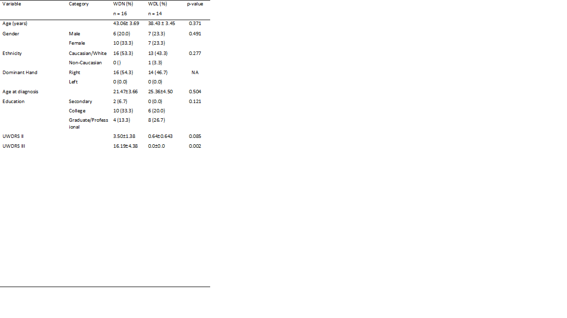
Method: Subjects (n=30) with treated WD were recruited from the WD clinic at Yale. Those with hepatic encephalopathy or active complications of portal hypertension were excluded. Neurologic disease severity was assessed using the Unified Wilson’s Disease Rating Scale (UWDRS). Subjects with a UWDRS part III (motor) score greater than 0 were assigned to the neurologic (WDN) group, while those with a score of 0 were assigned to the liver (WDL) group. Control subjects were age and gender-matched from a Yale imaging database. T1-weighted MPRAGE anatomical images were collected in a 3T scanner. Cortical and subcortical volumetric analysis was performed using the automated segmentation algorithm in the FreeSurfer software (1). Based on our a priori hypothesis, we focused on volumetric differences of the thalamus and basal ganglia (normalized by the total intracranial volume) between WDN, WDL, and control groups.
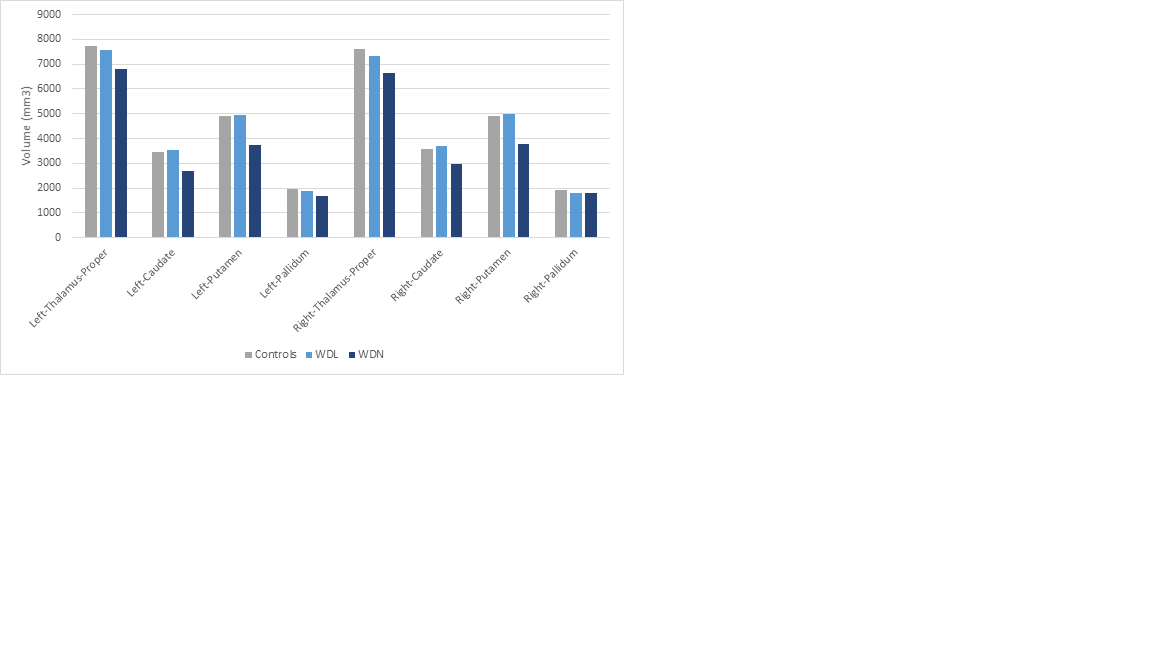
Results: WDN (n=16) and WDL (n=14) subjects were studied, with similar demographics and total brain volume. [table1] WDN subjects showed statistically significant volume loss of the thalamus and basal ganglia compared to age-matched controls. WDL subjects showed no atrophy of these structures. [figure1] Both groups had similar total gray matter volume. Cerebral white matter and ventricle size was reduced in both WDN and WDL groups.
Conclusion: Structural changes, namely atrophy of the thalamus and basal ganglia, can be quantified and are readily apparent in WDN patients. Purely hepatic phenotype WD subjects still demonstrate non-specific white matter loss and ventricular enlargement. These changes may be future areas of neuroimaging study in neurologically-intact WD patients.
References: 1. Fischl, B., Salat, D.H., Busa, E., Albert, M., Dieterich, M., Haselgrove, C., van der Kouwe, A., Killiany, R., Kennedy, D., Klaveness, S., Montillo, A., Makris, N., Rosen, B., Dale, A.M., 2002. Whole brain segmentation: automated labeling of neuroanatomical structures in the human brain. Neuron 33, 341-355.
To cite this abstract in AMA style:
A. Patel, K. Nalamada, A. Vives-Rodriguez, D. Robakis, T. Constable, J. Arora, M. Schilsky, S. Tinaz. Structural imaging changes in hepatic and neurological Wilson disease [abstract]. Mov Disord. 2019; 34 (suppl 2). https://www.mdsabstracts.org/abstract/structural-imaging-changes-in-hepatic-and-neurological-wilson-disease/. Accessed March 21, 2026.« Back to 2019 International Congress
MDS Abstracts - https://www.mdsabstracts.org/abstract/structural-imaging-changes-in-hepatic-and-neurological-wilson-disease/