Objective: The purpose of this study was to explore the diagnostic value of PET imaging, plasma biomarkers in the diagnosis of cognitive dysfunction in Parkinson’s disease(PD), including subjective cognitive decline (SCD), mild cognitive impairment (MCI) and PD dementia (PDD).
Background: Cognitive impairment is a major complication in Parkinson’s disease(PD). The clinical spectrum of PD-related cognitive impairment includes SCD,MCI and PDD. Since identifying cognitive decline at its earliest stage in PD may improve patient management, there is increasingly interest in evaluating the value of SCD in PD as an initial marker of cognitive change.
Method: A total of 147 patients with PD were enrolled in this study. Based on cognitive function assessments, patients were categorized into four groups: PD‐NC(n = 34),PD‐SCD(n = 33) ,PD‐MCI( n = 57), and PDD( n = 23). Baseline clinical data were collected, and all patients underwent dual positron emission tomography (PET) imaging using DAT‐PET—with either DTBZ or CFT as the imaging agent—and FDG‐PET to assess brain imaging characteristics associated with cognitive function in PD.
Results: No significant differences in DAT‐PET uptake were observed among the PD cognitive groups. In 18F‐FDG PET imaging, the PDD group exhibited reduced metabolism in the precuneus, hippocampus, middle frontal gyrus, middle temporal gyrus, caudate nucleus, and left middle occipital gyrus compared with the PD‐MCI group. Furthermore, the PD‐MCI group showed significantly decreased metabolism in the superior temporal gyrus relative to the PD‐NC group, whereas the PD‐SCD group demonstrated increased metabolism in the bilateral nucleus accumbens and decreased metabolism in the bilateral inferior occipital gyrus compared with the PD‐NC group. Moreover, compared with the PD‐SCD group, the PD‐MCI group had reduced metabolism in the precuneus, middle cingulate gyrus, red nucleus, dorsomedial thalamic nucleus, and medial thalamic plate.
Conclusion: PD-MCI and PDD showed impairments in several cognitive functions, including visuospatial, executive, memory, language, and attention/working memory cognitive domains at an early stage. DAT-PET did not differ significantly in patients with each cognitive state of PD, while 18F-FDG PET imaging showed significant decreases in cortical metabolism in the frontal, temporal, and occipital lobes in the PDD group compared with other groups
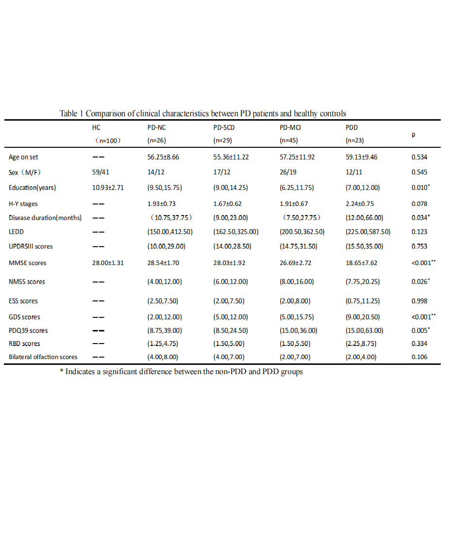
table1
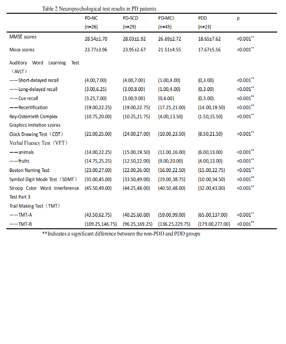
table2
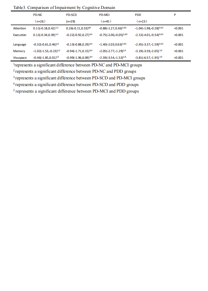
table3
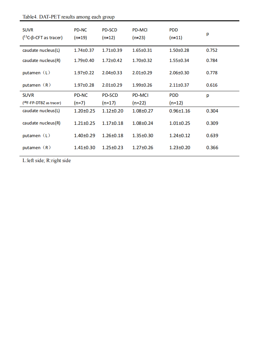
table4
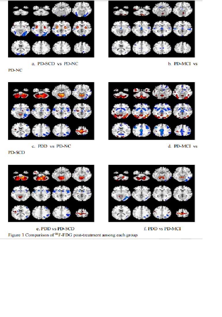
fig1
References: [1] FOLTYNIE T, BRAYNE C E, ROBBINS T W, et al. The cognitive ability of an incident cohort of Parkinson’s patients in the UK. The CamPaIGN study [J]. Brain : a journal of
neurology, 2004, 127(Pt 3): 550-60.
[2] WEINTRAUB D, MARRAS C, AMARA A, et al. Association between Subjective Cognitive Complaints and Incident Functional Impairment in Parkinson’s Disease [J]. Movement
disorders : official journal of the Movement Disorder Society, 2024, 39(4): 706-14.
To cite this abstract in AMA style:
T. Hu, Y. Tang, J. Wang. The value of PET imaging in Parkinson’s disease with cognition impairment [abstract]. Mov Disord. 2025; 40 (suppl 1). https://www.mdsabstracts.org/abstract/the-value-of-pet-imaging-in-parkinsons-disease-with-cognition-impairment/. Accessed April 10, 2026.« Back to 2025 International Congress
MDS Abstracts - https://www.mdsabstracts.org/abstract/the-value-of-pet-imaging-in-parkinsons-disease-with-cognition-impairment/