Category: Parkinson's disease: Neuroimaging
Objective: To present the usage and results of transcranial sonography (TCS) in Parkinson’s disease (PD) patients.
Background: (TCS) has emerged as a valuable diagnostic tool in movement disorder centers worldwide, particularly for Parkinson’s disease (PD). Its non-invasive nature, cost-effectiveness, and ability to provide unique insights into brain structures make it a preferred method for both clinical and research purposes. TCS has gained widespread acceptance in movement disorder centers due to its ability to detect specific sonographic features associated with PD. The most notable finding is the hyperechogenicity (HE) of the substantia nigra (SN), observed in over 90% of idiopathic PD patients [1-3].
Method: The results obtained from the routine neurological assessment in the Republican Centre for Movement Disorders (Kazan, Russia)
Results: The study included 201 participants, comprising 92 relatively healthy controls and 109 patients with PD. TCS was performed to assess HE in the SN. In the control group, 8 individuals (8.7%) exhibited HE, with 2 (2.2%) showing pathologically increased SN HE (exceeding 20 mm²). In contrast, 30 PD patients (27.5%) demonstrated HE, with 5 (4.6%) having values below the 20 mm² threshold (Fig. 1).
The numerical features of SN HE are detailed in Table 1, while the correlation between the sizes of HE in the right and left SN is illustrated in Fig. 2. Logistic regression analysis was conducted to evaluate the predictive value of pathological TCS findings for PD. The model was statistically significant (Chi²(1)=20.95, p<.001, n=197). The coefficient for pathological TCS (b=2.55) indicated a positive association, meaning that the presence of pathological TCS findings significantly increased the likelihood of PD. The odds ratio of 12.8 (p=.001) demonstrated that individuals with pathological TCS findings were 12.8 times more likely to have PD compared to those without such findings.
Conclusion: The findings highlight the diagnostic utility of TCS in differentiating PD patients from healthy controls. Pathological SN HE, particularly values exceeding 20 mm², was strongly associated with PD. This suggests that TCS can serve as a valuable tool in the early detection and diagnosis of PD, especially when combined with clinical evaluation. However, the presence of HE below the 20 mm² threshold in some PD patients indicates the need for further research to refine diagnostic criteria and improve the accuracy of TCS in PD diagnosis.
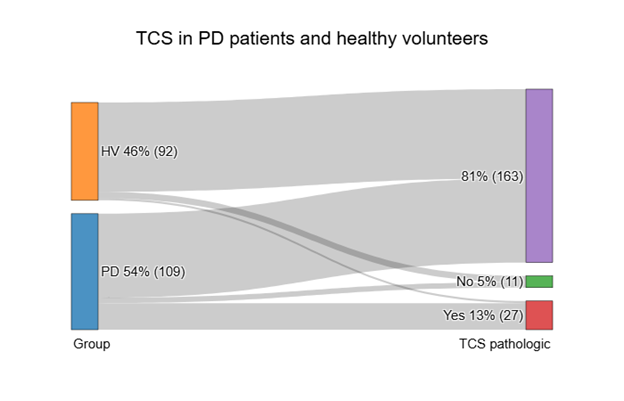
Figure 1. TCS frequency
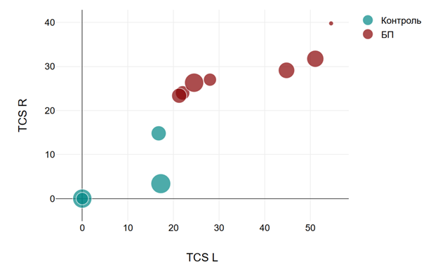
Figure 2. TCS right and left
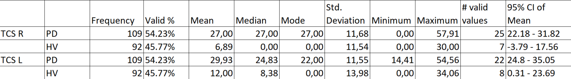
Table 1. Descriptive statistics
References: 1. Behnke, S., & Berg, D. (2013). Transcranial Ultrasonography in Movement Disorders (pp. 71–92). Humana Press, Totowa, NJ. https://doi.org/10.1007/978-1-62703-471-5_5
2. Berg, D., Godau, J., & Walter, U. (2008). Transcranial sonography in movement disorders. Lancet Neurology, 7(11), 1044–1055. https://doi.org/10.1016/S1474-4422(08)70239-4
3. Moskalenko, A. N., Chechetkin, A., Filatov, A., Fedotova, E. Yu., Konovalov, R. N., & Illarioshkin, S. (2023). Clinical and neuroimaging study of patients with Parkinson’s disease using transcranial sonography and neuromelanin-sensitive magnetic resonance imaging. Rossijskij Nevrologičeskij Žurnal, 27(6), 32–40. https://doi.org/10.30629/2658-7947-2022-27-6-32-40
To cite this abstract in AMA style:
D. Khasanova, I. Khasanov, Z. Zalyalova. Transcranial sonography in Parkinson’s disease patients [abstract]. Mov Disord. 2025; 40 (suppl 1). https://www.mdsabstracts.org/abstract/transcranial-sonography-in-parkinsons-disease-patients/. Accessed April 10, 2026.« Back to 2025 International Congress
MDS Abstracts - https://www.mdsabstracts.org/abstract/transcranial-sonography-in-parkinsons-disease-patients/