Session Information
Date: Wednesday, June 7, 2017
Session Title: Neuroimaging (Non-PD)
Session Time: 1:15pm-2:45pm
Location: Exhibit Hall C
Objective: To identify MRI-based brain networks of cerebral perfusion and structural abnormalities in cerebellar variant of MSA.
Background: MSA consists clinically of parkinsonian (MSA-P) and cerebellar (MSA-C) variants. Previous FDG PET studies have showed that MSA is associated with a specific disease-related pattern (MSARP) of cerebral glucose metabolism and its expression in individual patients could aid differential diagnosis of parkinsonism and predict clinical disability and disease duration (1). It is currently not known whether MRI modality can be used to generate analogous brain networks corresponding to different variants of MSA.
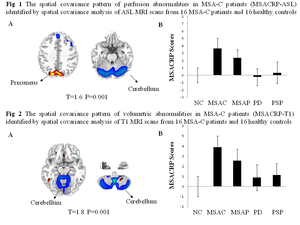
Methods: 16 Chinese patients with MSA-C (12M/4F, age 57.3 ± 6.7y), together with 16 MSA-P (10M/6F, age 59.6±8.2y), 16 PD (12M/4F, age 58.4±4.5y), 16 PSP (12M/4F, age 62.9±9.0y) and 16 healthy controls (6M/10F, age 58.7±4.6y) underwent arterial spin labeling (ASL) perfusion MRI and T1 structural MRI at 3 Tesla. SSMPCA toolbox was used with MSA-C patient and control images to identify brain networks associated with perfusion and volumetric changes (2). Each network was determined in a logistical regression model such that its expression best discriminated patients from controls with the lowest Akaike information criterion (AIC) value. Network expression was prospectively computed for other patients and compared across the groups.
Results: MSA-C perfusion pattern (MSACRP-ASL; 45% subject x voxel variance) was characterized bilaterally by decreases in the cerebellum and increases in the precuneus (Fig 1A). Its expression was elevated in the MSA-C and MSA-P patients (p<0.0001; Fig 1B) compared with the controls and PD/PSP patients but lower in the MSA-P than in MSA-C patients (p<0.01). MSA-C volume pattern (MSACRP-T1; 33% subject x voxel variance) was characterized by brain atrophy in the bilateral cerebellum (Fig 2A). Its expression was increased relative to the healthy controls in the MSA-C and MSA-P patients (p<0.0001; Fig 2B) and in the PD (p<0.05) and PSP (p<0.005) patients. The elevation was greater in the MSA-C than in MSA-P patients with both MSA groups higher than the PD and PSP patients (p<0.002).
Conclusions: MSA-C patients presented distinct brain networks of abnormal cerebral perfusion and atrophy. Both measures may provide viable markers to discriminate two clinical subtypes of MSA as well as in differential diagnosis of parkinsonism.
References: 1. Poston, KL (2012). Network correlates of disease severity in multiple system atrophy. Neurology 78: 1237-1244
2. Peng, S (2014). Characterization of disease-related covariance topographies with SSMPCA toolbox: effects of spatial normalization and PET scanners. Hum Brain Mapp 35: 1801-14
To cite this abstract in AMA style:
P. Wu, S. Peng, J. Ge, J. Wang, D. Eidelberg, C. Zuo, Y. Ma. MRI-based Brain Networks of Perfusion and Structural Abnormalities in Patients with Cerebellar Variant of Multiple System Atrophy [abstract]. Mov Disord. 2017; 32 (suppl 2). https://www.mdsabstracts.org/abstract/mri-based-brain-networks-of-perfusion-and-structural-abnormalities-in-patients-with-cerebellar-variant-of-multiple-system-atrophy/. Accessed June 28, 2026.« Back to 2017 International Congress
MDS Abstracts - https://www.mdsabstracts.org/abstract/mri-based-brain-networks-of-perfusion-and-structural-abnormalities-in-patients-with-cerebellar-variant-of-multiple-system-atrophy/