Category: Neuroimaging (Non-PD)
Objective: To describe MRI signs suggestive of dentatorubral-pallidoluysian atrophy (DRPLA) that may warrant genetic testing.
Background: Dentatorubral-pallidoluysian atrophy (DRPLA) is a rare autosomal dominant neurodegenerative disorder with a highly heterogeneous presentation that may differ with age of onset (genetic anticipation and CAG repetition length). It is characterized by symptoms such as epilepsy, myoclonus, cerebellar ataxia, choreoathetosis and dementia [1].
DRPLA is most prevalent in Japan but has also been described in non-Asian population. In fact, it is the second most common genetic cause of ataxia in Portugal.
Diagnosis is made by genetic testing, but MRI is usually done at an earlier stage of investigation and some changes may suggest the diagnosis.
Method: Clinical case description and brief literature review.
Results: A 20-year-old male presented with epilepsy, ataxia and scanning speech, as well as prior reported delayed developmental milestones during early childhood. His father had DRPLA genetically confirmed.
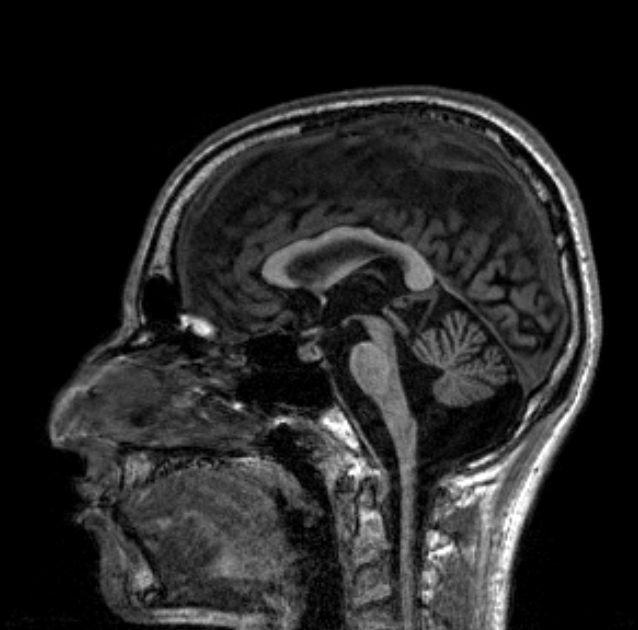
Brain MRI revealed cerebral atrophy, especially in infratentorial regions, with atrophy of both cerebellar hemispheres, and, more markedly, of the vermis. Additionally, the midsagittal image clearly showed a characteristic volume reduction of pontine tegmentum [figure 1].
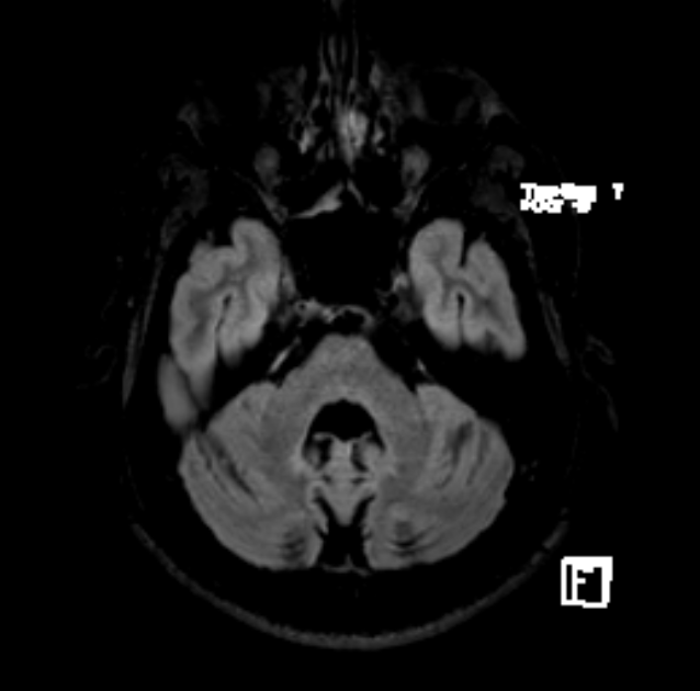
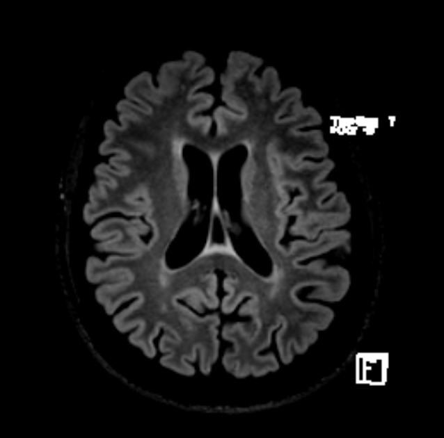
On T2/FLAIR there was white matter hyperintensity around the fourth ventricle and reaching the dentate nuclei [figure 2]. There were also linear periventricular hyperintensity in supratentorial compartment and scarce high-signal-intensity foci distributed on fronto-parietal region bilaterally [figure 3].
Conclusion: Atrophic changes in the cerebellum and, particularly, in the pontine tegmentum are typical MRI features of DRPLA [2]. Marked progression of cerebral atrophy is often noted in patients with juvenile-onset, and cerebral white matter is usually involved in an advanced stage.
Although genetic testing is essential for the diagnosis of DRPLA, common MRI features may raise awareness to this rare disease, even at an early stage.
References: 1. Veneziano L, Frontali M. DRPLA. 1999 Aug 6 [Updated 2016 Jun 9]. In: Adam MP, Ardinger HH, Pagon RA, et al., editors. GeneReviews® [Internet]. Seattle (WA): University of Washington, Seattle; 1993-2019
2. Sunami Y, Koide R, Arai N, et al. Radiologic and neuropathologic findings in patients in a family with dentatorubral-pallidoluysian atrophy. AJNR Am J Neuroradiol 2011;32:109–14
To cite this abstract in AMA style:
H. Queirós, I. Carneiro, E. Martins, A. Morgadinho, D. Pereira, G. Cordeiro. MR imaging features of Dentatorubral-pallidoluysian atrophy (DRPLA): a case report [abstract]. Mov Disord. 2022; 37 (suppl 2). https://www.mdsabstracts.org/abstract/mr-imaging-features-of-dentatorubral-pallidoluysian-atrophy-drpla-a-case-report/. Accessed June 24, 2026.« Back to 2022 International Congress
MDS Abstracts - https://www.mdsabstracts.org/abstract/mr-imaging-features-of-dentatorubral-pallidoluysian-atrophy-drpla-a-case-report/