Category: Parkinson's Disease: Neuroimaging
Objective: We utilized neurite orientation dispersion and density imaging (NODDI) along with diffusion tensor and kurtosis imaging (DTI/DKI) to assess brainstem nuclei in Parkinson’s disease subtypes that are involved in gait, posture, and motor control, including the pontomedullary reticular formation (pmRF), pedunculopontine nuclei (PPN) and the dorsal raphe (DR).
Background: NODDI is an advanced MRI technique that involves a three-compartmental voxel model and estimates the microstructural environment of neurites and extra-neurite space. DTI and its extension, DKI are also MR techniques and use multiple B values to measure the diffusion of water in a Gaussian and non-Gaussian distribution, respectively.
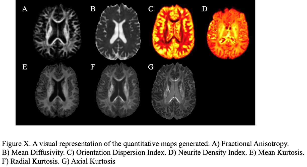
Method: 43 PD patients were analyzed with the subtypes: gait disturbance without freezing of gait (GD), freezing of gait (FOG), and non-gait disturbance (ND) and seven quantitative indices were obtained. These included (1) orientation dispersion index (ODI) and intracellular volume fraction (ICVF) derived from NODDI modeling; (2) fractional anisotropy (FA) and mean diffusivity (MD) computed from DTI and (3) mean kurtosis (MK), axial kurtosis (AK) and radial kurtosis (RK) from DKI. Unified Parkinson’s Disease Rating Scale (MDS-UPDRS) scores were obtained. ROIs (pmRF, PPN and DR) were selected from AAL atlas, manually revised and transferred to each patient diffusion MRI space using a nonlinear registration technique. Averaged diffusion quantitative values were computed. Analyses included correlation with UPDRS III scores using Pearson correlation and pairwise comparison with respect to the GD using ANOVA.
Results: The ODI in the pmRF had a strong positive correlation with the UPDRS in the TD group (p=0.009, r= 0.84). There was a strong negative correlation of the FA with the UPDRS in the TD group (p=0.004, r= -0.87). In TD group, there was a strong positive correlation with the UPDRS in the DR and left PPN (p=0.029, r= 0.76 and p=0.042, r=0.73, respectively). MD for the left pmRF was lower in the FOG group compared to GD (p=0.0411). ICVF for the left pmRF was lower in the GD compared to FOG (p=0.0221).
Conclusion: Correlations of the brainstem nuclei using NODDI and DTI/DKI with UPDRS seem to implicate differential involvement of these regions in PD subtypes. Further analysis of subcortical nuclei along with tractography may help delineate interactions between these regions.
To cite this abstract in AMA style:
N. Joshi, I. Aisles, I. Shelley, K. Talekar, M. Alizadeh. Neurite Orientation Dispersion and Density Imaging and Diffusion Kurtosis Imaging in Brainstem Nuclei for Subtypes of Parkinson’s Disease [abstract]. Mov Disord. 2023; 38 (suppl 1). https://www.mdsabstracts.org/abstract/neurite-orientation-dispersion-and-density-imaging-and-diffusion-kurtosis-imaging-in-brainstem-nuclei-for-subtypes-of-parkinsons-disease/. Accessed June 20, 2026.« Back to 2023 International Congress
MDS Abstracts - https://www.mdsabstracts.org/abstract/neurite-orientation-dispersion-and-density-imaging-and-diffusion-kurtosis-imaging-in-brainstem-nuclei-for-subtypes-of-parkinsons-disease/