Category: MSA, PSP, CBS: Neuroimaging
Objective: To demonstrate the overlap on radiographic metrics between NPH and PSP.
Background: The neuroradiological hallmark of NPH is non-obstructive ventriculomegaly (Evans’ index (EI) >0.3). Other features suggestive of NPH include an acute callosal angle, bowing of the corpus callosum, cingulate sulcus sign, and dilated Sylvian fissures with gyral crowding (disproportionately enlarged subarachnoid space hydrocephalus (DESH)). However, our preliminary analyses have shown that ventriculomegaly and other NPH-like imaging features may also be seen in PSP patients, causing uncertainty in diagnosis (Figure 1).
Method: We conducted a retrospective analysis on randomly selected patients with PSP (n=13) and compared them patients with NPH (n=29). For both cohorts, we obtained and compared the following radiographic metrics associated with NPH:
– Quantitative metrics: EIs, callosal angles, anteroposterior diameter of the lateral ventricle indices (ALVIs) (ventriculomegaly cutoff of 0.5)
– Qualitative metrics: DESH, cingulate sulcus sign, bowing of the corpus callosum.
Results: PSP patients had a mean EI [0.315 (vs. 0.362 in NPH), (p=0.261)] and ALVI [0.514 (vs. 0.595 in NPH), (p=0.191)] above the threshold for ventriculomegaly. Moreover, the PSP cohort demonstrated a smaller mean callosal angle than the NPH cohort (116.2° vs. 121.2°, respectively (p=0.0093)).
On qualitative metrics, DESH was demonstrated in 46.1% of PSP patients (vs. 82.7% of NPH patients, p=0.015). Cingulate sulcus sign was demonstrated in 7.7% of PSP patients (vs. 10.3% of NPH patients, p=0.786). Bowing of the corpus callosum was seen in 7.7% of PSP patients (vs. 37.4% of NPH patients, p=0.045).
Conclusion: PSP patients may exhibit several imaging features characteristic of NPH. While certain qualitative metrics may be more common in NPH, the prevalence of NPH-like features in PSP patients demonstrated the need for caution when interpreting imaging and allocating a presumptive NPH diagnosis, due to the risk of subjecting patients to unnecessary spinal taps and shunt surgeries (with their associated complications) when they instead may have PSP.
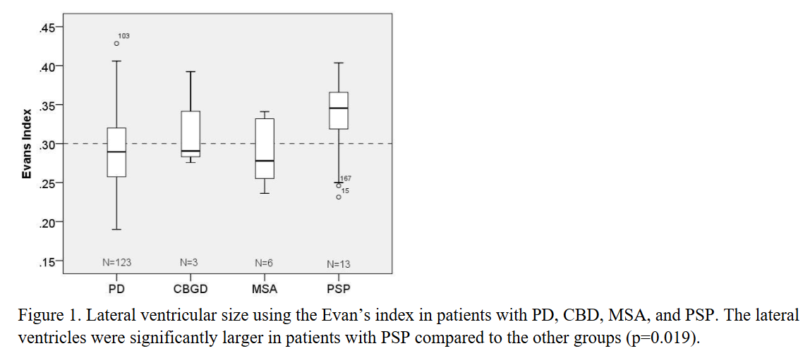
Figure 1
References: Önder H, Kocer B, Turan A, Kertmen H, Comoglu S. The Overlap in Neuroimaging Findings Between Idiopathic Normal Pressure Hydrocephalus and Progressive Supranuclear Palsy. Ann Indian Acad Neurol. 2022;25(6):1087-1091. doi/full/10.1111/jon.13204
Martin C, Schwartz G, Reinsel R. Radiologic Overlap between Idiopathic Normal Pressure Hydrocephalus and the Parkinsonian Syndromes. Neurology, 94(15_supplement).2020; 960.
Fu MH, Huang CC, Wu KLH, et al. Higher prevalence of idiopathic normal pressure hydrocephalus-like MRI features in progressive supranuclear palsy: An imaging reminder of atypical parkinsonism. Brain Behav. 2023;13(2):e2884. doi:10.1002/brb3.2884
To cite this abstract in AMA style:
R. Mani, C. Pol, M. Egnor, G. Schwartz. The Radiographic Overlap Between Normal Pressure Hydrocephalus (NPH) and Progressive Supranuclear Palsy (PSP): When Looks Can Be Deceiving [abstract]. Mov Disord. 2025; 40 (suppl 1). https://www.mdsabstracts.org/abstract/the-radiographic-overlap-between-normal-pressure-hydrocephalus-nph-and-progressive-supranuclear-palsy-psp-when-looks-can-be-deceiving/. Accessed July 6, 2026.« Back to 2025 International Congress
MDS Abstracts - https://www.mdsabstracts.org/abstract/the-radiographic-overlap-between-normal-pressure-hydrocephalus-nph-and-progressive-supranuclear-palsy-psp-when-looks-can-be-deceiving/