Category: Tremor
Objective: We performed the electrophysiological analysis of patients with MRI-confirmed hypertrophic olivary degeneration by surface and needle EMG.
Background: Hypertrophic olivary degeneration is a multisynaptic and transneuronal degeneration of inferior olivary nucleus triggered by deafferentation in the dentato-rubro-olivary pathway. Pathologically, the increased volume consists of cytoplasmic vacuolar degeneration and an increase in astrocyte number. The presentation includes Holmes tremor (HT), palatal tremor (PT), cerebellar ataxia, and ocular tremor.
There have been only 2 case reports in electrophysiology. One showed the surface EMG bursting frequency at 3Hz and bursting duration of 130ms in the oribularis oris muscle. The other showed bursting frequency at 2.2 Hz in the masseter muscles. Complete neurophysiology study is lacking. Thus we performed and analyzed surface and needle EMG on patients with various clinical presentations.
Method: From 2016 to 2024, 4 patients with MRI-confirmed inferior olivary nucleus hypertrophy were recruited. Surface EMG of the twitching muscles, needle EMG of the palate, and EEG were recorded. The signal analysis was performed by R-4.4.3 for Windows.
Results: All patients with inferior olivary nucleus hypertrophy had contralateral cerebellar ataxia while only 3 out of 4 had palatal tremor. Paramedian dorsal pons lesion were observed in 2 patients, one developed bilateral inferior olivary hypertrophy with bilateral palatal tremor while the other developed contralateral inferior olivary hypertrophy without palatal tremor [table].
Electrophysiologically, all patients exhibited well-formed and 150-200ms bursting activity in all sampled muscles, including those of the palate, face, and upper and lower limbs. There was a periodic wax-and-wane pattern of the bursting amplitudes at a fixed frequency ranging from 1 to 3 Hz [figure]. The time lag from palatal to facial muscle bursting was 25ms, and from palatal to shoulder muscle bursting was 50ms.
Conclusion: Contralateral cerebellar ataxia instead of palatal tremor is the most common sign of hypertrophic olivary degeneration. There are common electrophysiological features of 150-200ms duration, well-forming bursting pattern, and wax-and-wane amplitudes across all involved muscles. Our finding supports the hypothesis of dual mechanism: oscillations originating in the hypertrophic inferior olive and amplified by the cerebellum cortex.
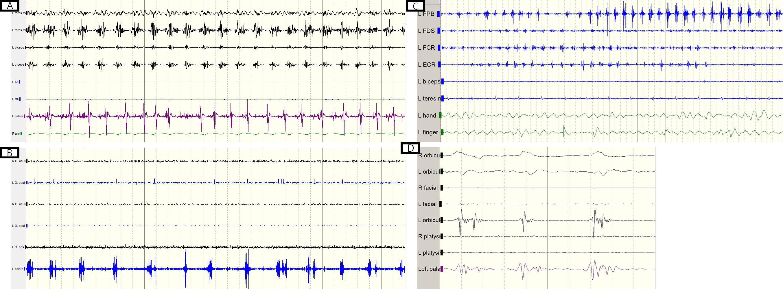
Figure
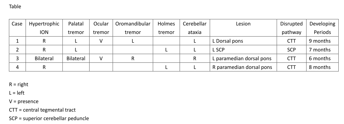
Table
References: 1. Wang H, Wang Y, Wang R, et al. Hypertrophic olivary degeneration: A comprehensive review focusing on etiology. Brain Res 2019;1718:53-63.
2. Goto N, Kaneko M. Olivary enlargement: chronological and morphometric analyses. Acta Neuropathol 1981;54:275-282.
3. Sanchez Hernandez J, Paniagua Escudero JC, Carreno Moran P, Asensio Calle JF. Hypertrophic olivary degeneration secondary to a Guillain-Mollaret triangle lesion. Neurologia 2013;28:59-61.
4. Martins WA, Schilling LP, Neto FK, Becker J. Hypertrophic Olivary Degeneration Secondary to Neuro-Behcet’s Disease. Clin Neuroradiol 2016;26:99-102.
5. Assenza F, Genovese M, Cabboi MP, Salomone G, Cavallieri F, Valzania F. Peribuccal and pharyngeal myorhythmia as a presenting symptom of hypertrophic olivary degeneration. Parkinsonism Relat Disord 2021;85:141-143.
6. Fernandez J, Garcia-Garcia A, Valle N, Infante J. Oculomandibular Tremor and Bilateral Hypertrophic Olivary Degeneration. Mov Disord Clin Pract 2017;4:152-153.
7. Shaikh AG, Hong S, Liao K, et al. Oculopalatal tremor explained by a model of inferior olivary hypertrophy and cerebellar plasticity. Brain 2010;133:923-940.
To cite this abstract in AMA style:
YCT. Tai, TKL. Lin. Clinical and Electrophysiological Features of Hypertrophic Olivary Degeneration [abstract]. Mov Disord. 2025; 40 (suppl 1). https://www.mdsabstracts.org/abstract/clinical-and-electrophysiological-features-of-hypertrophic-olivary-degeneration/. Accessed July 10, 2026.« Back to 2025 International Congress
MDS Abstracts - https://www.mdsabstracts.org/abstract/clinical-and-electrophysiological-features-of-hypertrophic-olivary-degeneration/