Category: Non-Dystonia (Other)
Objective: To describe a case of left facial synkinesis in a patient with a right middle cerebral artery stroke.
Background: Facial synkinesis has been classically known to be due to misguided nerve regeneration after axonal damage. This can happen after Bell’s palsy or surgical procedures that can injure the facial nerve. Even though this is the most widely accepted mechanism, it has been suggested that higher cortical reorganization can contribute to facial synkinesis [1]. This has been supported by studies showing activation of the supplementary motor area in patients with ipsilateral synkinesis [2]. This is a case of a patient with no history or electrodiagnostic evidence of facial nerve injury who developed synkinesis after a contralateral right middle cerebral artery stroke.
Method: Case report.
Results: A 59-year-old woman with a past medical history of hypertension, hyperlipidemia, atrial fibrillation, and endocarditis presented to the hospital due to sudden-onset left-sided weakness. A brain MRI showed a right middle cerebral artery stroke. A month after this, the patient returned to the hospital for abnormal movements of her left face and concern for possible focal status epilepticus. An EEG did not report any epileptiform activity. On exam, the patient had left-sided upper motor neuron weakness involving her face, arm, and leg. She was able to raise her eyebrows and close her eyes symmetrically. With closer observation, it was noted that the left side of her mouth would activate every time the patient blinked, consistent with synkinesis. There were no signs of hemifacial spasm. The patient and her family members denied any history of facial nerve involvement before the stroke. Her brain MRI was reviewed, and there was no pathology involving her brainstem. An EMG surveying the bilateral nasalis, orbicularis oculi, and orbicularis oris muscles showed normal amplitudes. Bilateral trigeminal blink responses showed symmetric R1 latencies, findings not suggestive of left-sided facial neuropathy.
Conclusion: Synkinesis is a movement disorder that can have functional and emotional consequences in our patients. Understanding possible alternative mechanisms for its pathogenesis can lead to the development of additional rehabilitation techniques, including stimulation of cortical neural networks that can get involved. Further studies, including transcranial magnetic stimulation [2], are recommended to investigate its pathophysiology.
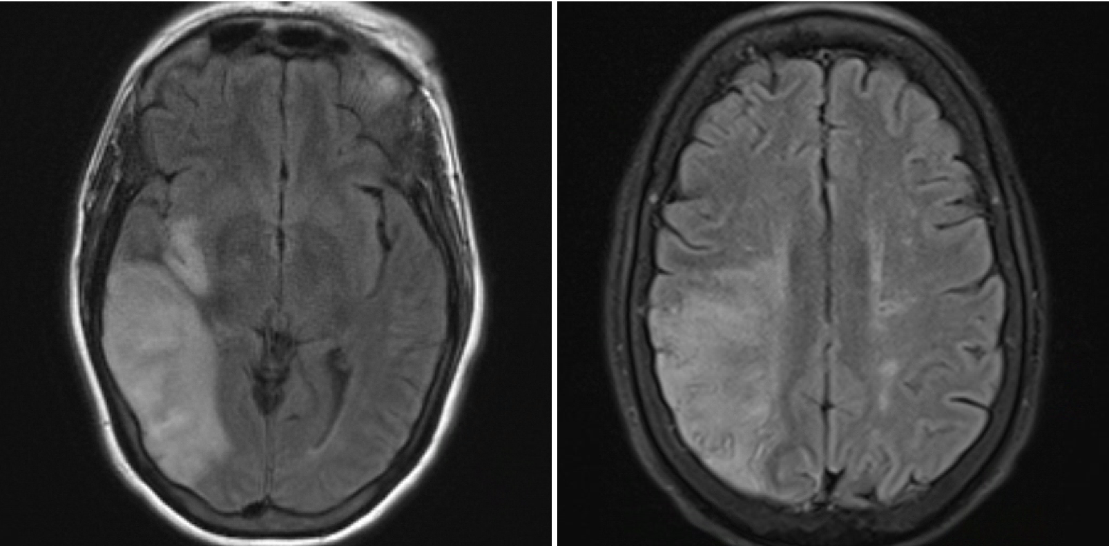
Figure 1. Brain MRI

A. Smiling; B. Closing eyes; C. Both tasks
References: 1. Guntinas-Lichius O, Prengel J, Cohen O, Mäkitie AA, Vander Poorten V, Ronen O, Shaha A, Ferlito A. Pathogenesis, diagnosis and therapy of facial synkinesis: A systematic review and clinical practice recommendations by the international head and neck scientific group. Front Neurol. 2022 Nov 9;13:1019554. doi: 10.3389/fneur.2022.1019554. PMID: 36438936; PMCID: PMC9682287.
2. Salardini A, Narayanan NS, Arora J, Constable T, Jabbari B. Ipsilateral synkinesia involves the supplementary motor area. Neurosci Lett. 2012;523(2):135-138. doi:10.1016/j.neulet.2012.06.060.
To cite this abstract in AMA style:
J. Patino. Facial Synkinesis: Can It Have a Central Origin? [abstract]. Mov Disord. 2025; 40 (suppl 1). https://www.mdsabstracts.org/abstract/facial-synkinesis-can-it-have-a-central-origin/. Accessed July 10, 2026.« Back to 2025 International Congress
MDS Abstracts - https://www.mdsabstracts.org/abstract/facial-synkinesis-can-it-have-a-central-origin/